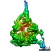
 Movie
Movie Controller

[English] 日本語
 Yorodumi
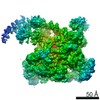
- PDB-6pb5: The E. coli class-II CAP-dependent transcription activation compl... -
+ Open data

- Basic information
| Entry | Database: PDB / ID: 6pb5 | ||||||
|---|---|---|---|---|---|---|---|
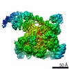
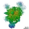
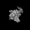
| Title | The E. coli class-II CAP-dependent transcription activation complex at the state 1 architecture | ||||||
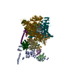
 Components Components |
| ||||||
 Keywords Keywords | TRANSCRIPTION/DNA / class-II / CAP-dependent / transcription activation complex / TRANSCRIPTION / TRANSCRIPTION-DNA complex | ||||||
| Function / homology |  Function and homology information Function and homology informationsigma factor antagonist complex / RNA polymerase complex / submerged biofilm formation / cellular response to cell envelope stress / regulation of DNA-templated transcription initiation / sigma factor activity / bacterial-type flagellum assembly / bacterial-type RNA polymerase core enzyme binding / cytosolic DNA-directed RNA polymerase complex / bacterial-type flagellum-dependent cell motility ...sigma factor antagonist complex / RNA polymerase complex / submerged biofilm formation / cellular response to cell envelope stress / regulation of DNA-templated transcription initiation / sigma factor activity / bacterial-type flagellum assembly / bacterial-type RNA polymerase core enzyme binding / cytosolic DNA-directed RNA polymerase complex / bacterial-type flagellum-dependent cell motility / nitrate assimilation / cAMP binding / regulation of DNA-templated transcription elongation / transcription elongation factor complex / DNA-directed RNA polymerase complex / cell motility / transcription antitermination / DNA-templated transcription initiation / protein-DNA complex / ribonucleoside binding / DNA-directed RNA polymerase / DNA-directed RNA polymerase activity / response to heat / protein-containing complex assembly / sequence-specific DNA binding / intracellular iron ion homeostasis / protein dimerization activity / transcription cis-regulatory region binding / DNA-binding transcription factor activity / negative regulation of DNA-templated transcription / regulation of DNA-templated transcription / positive regulation of DNA-templated transcription / magnesium ion binding / DNA-templated transcription / DNA binding / zinc ion binding / membrane / cytosol / cytoplasm Similarity search - Function | ||||||
| Biological species |  | ||||||
| Method | ELECTRON MICROSCOPY / single particle reconstruction / cryo EM / Resolution: 4.52 Å | ||||||
 Authors Authors | Liu, B. / Shi, W. | ||||||
 Citation Citation | Journal: PLoS Biol / Year: 2020 Title: Visualization of two architectures in class-II CAP-dependent transcription activation. Authors: Wei Shi / Yanan Jiang / Yibin Deng / Zigang Dong / Bin Liu /   Abstract: Transcription activation by cyclic AMP (cAMP) receptor protein (CAP) is the classic paradigm of transcription regulation in bacteria. CAP was suggested to activate transcription on class-II promoters ...Transcription activation by cyclic AMP (cAMP) receptor protein (CAP) is the classic paradigm of transcription regulation in bacteria. CAP was suggested to activate transcription on class-II promoters via a recruitment and isomerization mechanism. However, whether and how it modifies RNA polymerase (RNAP) to initiate transcription remains unclear. Here, we report cryo-electron microscopy (cryo-EM) structures of an intact Escherichia coli class-II CAP-dependent transcription activation complex (CAP-TAC) with and without de novo RNA transcript. The structures reveal two distinct architectures of TAC and raise the possibility that CAP binding may induce substantial conformational changes in all the subunits of RNAP and transiently widen the main cleft of RNAP to facilitate DNA promoter entering and formation of the initiation open complex. These structural changes vanish during further RNA transcript synthesis. The observations in this study may reveal a possible on-pathway intermediate and suggest a possibility that CAP activates transcription by inducing intermediate state, in addition to the previously proposed stabilization mechanism. | ||||||
| History |
|
- Structure visualization
| Movie |
Movie viewer |
|---|---|
| Structure viewer | Molecule: MolmilJmol/JSmol |
- Downloads & links
-Download
| PDBx/mmCIF format | 6pb5.cif.gz | 1.4 MB | Display | PDBx/mmCIF format |
|---|---|---|---|---|
| PDB format | pdb6pb5.ent.gz | 1.1 MB | Display | PDB format |
| PDBx/mmJSON format | 6pb5.json.gz | Tree view | PDBx/mmJSON format | |
| Others |  Other downloads Other downloads |
-Validation report
| Arichive directory | https://data.pdbj.org/pub/pdb/validation_reports/pb/6pb5ftp://data.pdbj.org/pub/pdb/validation_reports/pb/6pb5 | HTTPS FTP |
|---|
-Related structure data
| Related structure data | 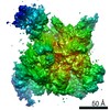 20287MC 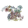 6pb4C 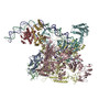 6pb6C M: map data used to model this data C: citing same article ( |
|---|---|
| Similar structure data |


-Links
 PDBj
PDBj


- Assembly
| Deposited unit | 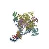
|
|---|---|
| 1 |
|
-Components
-DNA-directed RNA polymerase subunit ... , 4 types, 5 molecules ABCDE
| #1: Protein | Mass: 36558.680 Da / Num. of mol.: 2 Source method: isolated from a genetically manipulated source Source: (gene. exp.) References: UniProt: P0A7Z6, UniProt: P0A7Z4*PLUS, DNA-directed RNA polymerase #2: Protein | | Mass: 150820.875 Da / Num. of mol.: 1 Source method: isolated from a genetically manipulated source Source: (gene. exp.) #3: Protein | | Mass: 155366.781 Da / Num. of mol.: 1 Source method: isolated from a genetically manipulated source Source: (gene. exp.) #4: Protein | | Mass: 10249.547 Da / Num. of mol.: 1 Source method: isolated from a genetically manipulated source Source: (gene. exp.) References: UniProt: B7MFL0, UniProt: P0A800*PLUS, DNA-directed RNA polymerase |
|---|
-Protein , 2 types, 3 molecules FGH
| #5: Protein | Mass: 72206.266 Da / Num. of mol.: 1 Source method: isolated from a genetically manipulated source Source: (gene. exp.) |
|---|---|
| #6: Protein | Mass: 23672.439 Da / Num. of mol.: 2 Source method: isolated from a genetically manipulated source Source: (gene. exp.) |
-DNA chain , 2 types, 2 molecules 12
| #7: DNA chain | Mass: 23976.332 Da / Num. of mol.: 1 / Source method: obtained synthetically / Source: (synth.) |
|---|---|
| #8: DNA chain | Mass: 24191.590 Da / Num. of mol.: 1 / Source method: obtained synthetically / Source: (synth.) |
-Non-polymers , 3 types, 5 molecules 


| #9: Chemical |  Mass: 65.409 Da / Num. of mol.: 2 / Source method: obtained synthetically / Formula: Zn / Feature type: SUBJECT OF INVESTIGATION Mass: 65.409 Da / Num. of mol.: 2 / Source method: obtained synthetically / Formula: Zn / Feature type: SUBJECT OF INVESTIGATION#10: Chemical | ChemComp-MG / |  Mass: 24.305 Da / Num. of mol.: 1 / Source method: obtained synthetically / Formula: Mg / Feature type: SUBJECT OF INVESTIGATION Mass: 24.305 Da / Num. of mol.: 1 / Source method: obtained synthetically / Formula: Mg / Feature type: SUBJECT OF INVESTIGATION#11: Chemical |  Mass: 329.206 Da / Num. of mol.: 2 / Source method: obtained synthetically / Formula: C10H12N5O6P / Feature type: SUBJECT OF INVESTIGATION Mass: 329.206 Da / Num. of mol.: 2 / Source method: obtained synthetically / Formula: C10H12N5O6P / Feature type: SUBJECT OF INVESTIGATION |
|---|
-Details
| Has ligand of interest | Y |
|---|
-Experimental details
-Experiment
| Experiment | Method: ELECTRON MICROSCOPY |
|---|---|
| EM experiment | Aggregation state: PARTICLE / 3D reconstruction method: single particle reconstruction |
- Sample preparation
| Component | Name: The E coli class-II CAP-dependent transcription activation complex at the state 1 Type: COMPLEX / Entity ID: #1-#8 / Source: RECOMBINANT | ||||||||||||||||
|---|---|---|---|---|---|---|---|---|---|---|---|---|---|---|---|---|---|
| Molecular weight | Value: 0.55 MDa / Experimental value: NO | ||||||||||||||||
| Source (natural) | Organism: | ||||||||||||||||
| Source (recombinant) | Organism: | ||||||||||||||||
| Buffer solution | pH: 7.5 | ||||||||||||||||
| Buffer component |
| ||||||||||||||||
| Specimen | Conc.: 0.55 mg/ml / Embedding applied: NO / Shadowing applied: NO / Staining applied: NO / Vitrification applied: YES | ||||||||||||||||
| Specimen support | Details: 15 mA / Grid material: COPPER / Grid mesh size: 300 divisions/in. / Grid type: Quantifoil R1.2/1.3 | ||||||||||||||||
| Vitrification | Instrument: FEI VITROBOT MARK IV / Cryogen name: ETHANE / Humidity: 100 % / Chamber temperature: 277 K / Details: blot for 3 seconds before plunging |
- Electron microscopy imaging
| Experimental equipment |  Model: Titan Krios / Image courtesy: FEI Company |
|---|---|
| Microscopy | Model: FEI TITAN KRIOS |
| Electron gun | Electron source:  FIELD EMISSION GUN / Accelerating voltage: 300 kV / Illumination mode: FLOOD BEAM FIELD EMISSION GUN / Accelerating voltage: 300 kV / Illumination mode: FLOOD BEAM |
| Electron lens | Mode: BRIGHT FIELD / Nominal magnification: 96000 X / Nominal defocus max: 2600 nm / Nominal defocus min: 800 nm / Cs: 2.7 mm / C2 aperture diameter: 100 µm / Alignment procedure: COMA FREE |
| Specimen holder | Cryogen: NITROGEN / Specimen holder model: FEI TITAN KRIOS AUTOGRID HOLDER / Temperature (max): 77 K / Temperature (min): 77 K |
| Image recording | Average exposure time: 36 sec. / Electron dose: 36 e/Å2 / Detector mode: COUNTING / Film or detector model: FEI FALCON III (4k x 4k) / Num. of grids imaged: 1 / Num. of real images: 2819 |
- Processing
| EM software |
| |||||||||
|---|---|---|---|---|---|---|---|---|---|---|
| CTF correction | Type: NONE | |||||||||
| 3D reconstruction | Resolution: 4.52 Å / Resolution method: FSC 0.143 CUT-OFF / Num. of particles: 30296 / Symmetry type: POINT |
