 Movie
Movie Controller

[English] 日本語
 Yorodumi
- PDB-6p8u: Structure of P. aeruginosa ATCC27853 CdnD:HORMA2:Peptide 1 complex -
+ Open data

- Basic information
| Entry | Database: PDB / ID: 6p8u | ||||||
|---|---|---|---|---|---|---|---|
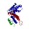
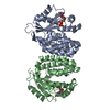
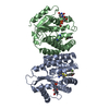
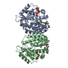
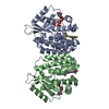
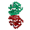
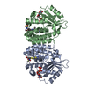
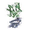
| Title | Structure of P. aeruginosa ATCC27853 CdnD:HORMA2:Peptide 1 complex | ||||||
 Components Components |
| ||||||
 Keywords Keywords | SIGNALING PROTEIN / second-messenger signaling / cGAS / CD-NTase / HORMA domain / closure motif | ||||||
| Function / homology |  Function and homology information Function and homology informationdiadenylate cyclase / diadenylate cyclase activity / nucleotide metabolic process / nucleotidyltransferase activity / Transferases; Transferring phosphorus-containing groups; Nucleotidyltransferases / defense response to virus / ATP binding / metal ion binding Similarity search - Function | ||||||
| Biological species |   Pseudomonas aeruginosa (bacteria) Pseudomonas aeruginosa (bacteria) | ||||||
| Method |  X-RAY DIFFRACTION / SYNCHROTRON / MOLECULAR REPLACEMENT / Resolution: 1.893 Å X-RAY DIFFRACTION / SYNCHROTRON / MOLECULAR REPLACEMENT / Resolution: 1.893 Å | ||||||
 Authors Authors | Ye, Q. / Corbett, K.D. | ||||||
 Citation Citation | Journal: Mol.Cell / Year: 2020 Title: HORMA Domain Proteins and a Trip13-like ATPase Regulate Bacterial cGAS-like Enzymes to Mediate Bacteriophage Immunity. Authors: Ye, Q. / Lau, R.K. / Mathews, I.T. / Birkholz, E.A. / Watrous, J.D. / Azimi, C.S. / Pogliano, J. / Jain, M. / Corbett, K.D. | ||||||
| History |
|
- Structure visualization
| Structure viewer | Molecule: MolmilJmol/JSmol |
|---|
- Downloads & links
-Download
| PDBx/mmCIF format | 6p8u.cif.gz | 283.5 KB | Display | PDBx/mmCIF format |
|---|---|---|---|---|
| PDB format | pdb6p8u.ent.gz | 231.4 KB | Display | PDB format |
| PDBx/mmJSON format | 6p8u.json.gz | Tree view | PDBx/mmJSON format | |
| Others |  Other downloads Other downloads |
-Validation report
| Arichive directory | https://data.pdbj.org/pub/pdb/validation_reports/p8/6p8uftp://data.pdbj.org/pub/pdb/validation_reports/p8/6p8u | HTTPS FTP |
|---|
-Related structure data
| Related structure data |  6p80C  6p82SC  6p8jC  6p8oC 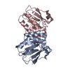 6p8pC 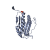 6p8rSC 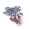 6p8sC  6p8vC  6pb3C  6u7bC S: Starting model for refinement C: citing same article ( |
|---|---|
| Similar structure data | |
| Experimental dataset #1 | Data set type: diffraction image data / Metadata reference: 10.15785/SBGRID/679 |


-Links
 PDBj
PDBj
- Assembly
| Deposited unit | 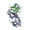
| ||||||||
|---|---|---|---|---|---|---|---|---|---|
| 1 |
| ||||||||
| Unit cell |
|
-Components
| #1: Protein | Mass: 34063.328 Da / Num. of mol.: 1 Source method: isolated from a genetically manipulated source Source: (gene. exp.) Pseudomonas aeruginosa (bacteria)Gene: DY979_07585, EGY23_20895, IPC669_24880, PA5486_02902, PAERUG_E15_London_28_01_14_04351, PAMH19_6112 Production host: |
|---|---|
| #2: Protein | Mass: 18723.082 Da / Num. of mol.: 1 Source method: isolated from a genetically manipulated source Source: (gene. exp.) Pseudomonas aeruginosa (bacteria)Gene: ORF C60, CAZ10_14260, DY940_15620, DY979_07580, EGY23_20890, EQH76_12140, IPC669_24875, PA5486_02901, PAERUG_E15_London_28_01_14_04350, PAMH19_6113 Production host: |
| #3: Protein/peptide | Mass: 1137.219 Da / Num. of mol.: 1 Source method: isolated from a genetically manipulated source Source: (gene. exp.) Pseudomonas aeruginosa (bacteria) / Production host: |
| #4: Chemical | ChemComp-MG /   Mass: 24.305 Da / Num. of mol.: 1 / Source method: obtained synthetically / Formula: Mg Mass: 24.305 Da / Num. of mol.: 1 / Source method: obtained synthetically / Formula: Mg |
| #5: Water | ChemComp-HOH /  Mass: 18.015 Da / Num. of mol.: 307 / Source method: isolated from a natural source / Formula: H2O Mass: 18.015 Da / Num. of mol.: 307 / Source method: isolated from a natural source / Formula: H2O |
| Has ligand of interest | N |
-Experimental details
-Experiment
| Experiment | Method: X-RAY DIFFRACTION / Number of used crystals: 1 |
|---|
- Sample preparation
| Crystal | Density Matthews: 2.08 Å3/Da / Density % sol: 40.98 % |
|---|---|
| Crystal grow | Temperature: 293 K / Method: vapor diffusion, hanging drop / pH: 5.5 Details: 100 mM Bis-Tris pH 5.5, 0.2 M ammonium acetate, and 30% PEG 3350 |
-Data collection
| Diffraction | Mean temperature: 100 K / Serial crystal experiment: N |
|---|---|
| Diffraction source | Source: SYNCHROTRON / Site: APS  / Beamline: 24-ID-C / Wavelength: 0.9791 Å / Beamline: 24-ID-C / Wavelength: 0.9791 Å |
| Detector | Type: DECTRIS PILATUS3 6M / Detector: PIXEL / Date: Mar 28, 2019 |
| Radiation | Protocol: SINGLE WAVELENGTH / Monochromatic (M) / Laue (L): M / Scattering type: x-ray |
| Radiation wavelength | Wavelength: 0.9791 Å / Relative weight: 1 |
| Reflection | Resolution: 1.89→81.97 Å / Num. obs: 34946 / % possible obs: 97.7 % / Redundancy: 3.3 % / CC1/2: 0.99 / Rmerge(I) obs: 0.134 / Rpim(I) all: 0.086 / Rrim(I) all: 0.159 / Net I/σ(I): 7.1 |
| Reflection shell | Resolution: 1.89→1.94 Å / Redundancy: 3.2 % / Rmerge(I) obs: 1.302 / Mean I/σ(I) obs: 0.8 / Num. unique obs: 1989 / CC1/2: 0.409 / Rpim(I) all: 0.837 / Rrim(I) all: 1.554 / % possible all: 87 |
- Processing
| Software |
| |||||||||||||||||||||||||||||||||||||||||||||||||||||||||||||||||||||||||||||||||||||||||||
|---|---|---|---|---|---|---|---|---|---|---|---|---|---|---|---|---|---|---|---|---|---|---|---|---|---|---|---|---|---|---|---|---|---|---|---|---|---|---|---|---|---|---|---|---|---|---|---|---|---|---|---|---|---|---|---|---|---|---|---|---|---|---|---|---|---|---|---|---|---|---|---|---|---|---|---|---|---|---|---|---|---|---|---|---|---|---|---|---|---|---|---|---|
| Refinement | Method to determine structure: MOLECULAR REPLACEMENT Starting model: 6P82, 6P8R Resolution: 1.893→81.966 Å / SU ML: 0.25 / Cross valid method: FREE R-VALUE / σ(F): 0.31 / Phase error: 24.39
| |||||||||||||||||||||||||||||||||||||||||||||||||||||||||||||||||||||||||||||||||||||||||||
| Solvent computation | Shrinkage radii: 0.9 Å / VDW probe radii: 1.11 Å | |||||||||||||||||||||||||||||||||||||||||||||||||||||||||||||||||||||||||||||||||||||||||||
| Refinement step | Cycle: LAST / Resolution: 1.893→81.966 Å
| |||||||||||||||||||||||||||||||||||||||||||||||||||||||||||||||||||||||||||||||||||||||||||
| Refine LS restraints |
| |||||||||||||||||||||||||||||||||||||||||||||||||||||||||||||||||||||||||||||||||||||||||||
| LS refinement shell |
| |||||||||||||||||||||||||||||||||||||||||||||||||||||||||||||||||||||||||||||||||||||||||||
| Refinement TLS params. | Method: refined / Refine-ID: X-RAY DIFFRACTION
| |||||||||||||||||||||||||||||||||||||||||||||||||||||||||||||||||||||||||||||||||||||||||||
| Refinement TLS group |
|
