 Movie
Movie Controller

[English] 日本語
 Yorodumi
- PDB-6p8l: Escherichia coli Bacterioferritin Substituted with Zinc Protoporp... -
+ Open data

- Basic information
| Entry | Database: PDB / ID: 6p8l | ||||||
|---|---|---|---|---|---|---|---|
| Title | Escherichia coli Bacterioferritin Substituted with Zinc Protoporphyrin IX (Zn Absorption Edge X-ray Data) | ||||||
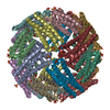
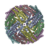
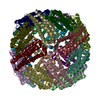
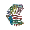
 Components Components | Bacterioferritin | ||||||
 Keywords Keywords | METAL BINDING PROTEIN / OXIDOREDUCTASE / bacterioferritin / zinc protoporphyrin IX / photosensitizer / ferritin | ||||||
| Function / homology |  Function and homology information Function and homology informationiron ion sequestering activity / ferroxidase / ferroxidase activity / ferric iron binding / iron ion transport / intracellular iron ion homeostasis / oxidoreductase activity / iron ion binding / heme binding / protein homodimerization activity ...iron ion sequestering activity / ferroxidase / ferroxidase activity / ferric iron binding / iron ion transport / intracellular iron ion homeostasis / oxidoreductase activity / iron ion binding / heme binding / protein homodimerization activity / membrane / identical protein binding / cytosol Similarity search - Function | ||||||
| Biological species |  | ||||||
| Method |  X-RAY DIFFRACTION / SYNCHROTRON / FOURIER SYNTHESIS / Resolution: 2.1 Å X-RAY DIFFRACTION / SYNCHROTRON / FOURIER SYNTHESIS / Resolution: 2.1 Å | ||||||
 Authors Authors | Taylor, A.B. / Cioloboc, D. / Kurtz, D.M. | ||||||
| Funding support |  United States, 1items United States, 1items
| ||||||
 Citation Citation | Journal: Biochemistry / Year: 2020 Title: Structure of a Zinc Porphyrin-Substituted Bacterioferritin and Photophysical Properties of Iron Reduction. Authors: Benavides, B.S. / Valandro, S. / Cioloboc, D. / Taylor, A.B. / Schanze, K.S. / Kurtz Jr., D.M. | ||||||
| History |
|
- Structure visualization
| Structure viewer | Molecule: MolmilJmol/JSmol |
|---|
- Downloads & links
-Download
| PDBx/mmCIF format | 6p8l.cif.gz | 484.8 KB | Display | PDBx/mmCIF format |
|---|---|---|---|---|
| PDB format | pdb6p8l.ent.gz | 370.7 KB | Display | PDB format |
| PDBx/mmJSON format | 6p8l.json.gz | Tree view | PDBx/mmJSON format | |
| Others |  Other downloads Other downloads |
-Validation report
| Arichive directory | https://data.pdbj.org/pub/pdb/validation_reports/p8/6p8lftp://data.pdbj.org/pub/pdb/validation_reports/p8/6p8l | HTTPS FTP |
|---|
-Related structure data
| Related structure data | 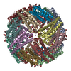 6p8kSC S: Starting model for refinement C: citing same article ( |
|---|---|
| Similar structure data |


-Links
 PDBj
PDBj


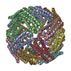
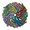
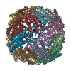
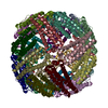
- Assembly
| Deposited unit | 
| |||||||||||||||
|---|---|---|---|---|---|---|---|---|---|---|---|---|---|---|---|---|
| 1 | 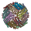
| |||||||||||||||
| Unit cell |
| |||||||||||||||
| Components on special symmetry positions |
|
-Components
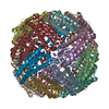
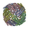
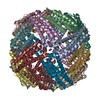
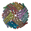
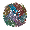
-Protein , 1 types, 12 molecules ABCDEFGHIJKL
| #1: Protein | Mass: 18518.016 Da / Num. of mol.: 12 Source method: isolated from a genetically manipulated source Source: (gene. exp.) |
|---|
-Non-polymers , 5 types, 1645 molecules 


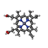

| #2: Chemical | ChemComp-ZN /  Mass: 65.409 Da / Num. of mol.: 12 / Source method: obtained synthetically / Formula: Zn Mass: 65.409 Da / Num. of mol.: 12 / Source method: obtained synthetically / Formula: Zn#3: Chemical | ChemComp-NA /  Mass: 22.990 Da / Num. of mol.: 4 / Source method: obtained synthetically / Formula: Na Mass: 22.990 Da / Num. of mol.: 4 / Source method: obtained synthetically / Formula: Na#4: Chemical | ChemComp-MLI /  Mass: 102.046 Da / Num. of mol.: 28 / Source method: obtained synthetically / Formula: C3H2O4 Mass: 102.046 Da / Num. of mol.: 28 / Source method: obtained synthetically / Formula: C3H2O4#5: Chemical | ChemComp-ZNH /  Mass: 626.051 Da / Num. of mol.: 6 / Source method: obtained synthetically / Formula: C34H32N4O4Zn / Feature type: SUBJECT OF INVESTIGATION Mass: 626.051 Da / Num. of mol.: 6 / Source method: obtained synthetically / Formula: C34H32N4O4Zn / Feature type: SUBJECT OF INVESTIGATION#6: Water | ChemComp-HOH / | Mass: 18.015 Da / Num. of mol.: 1595 / Source method: isolated from a natural source / Formula: H2O |
|---|
-Details
| Has ligand of interest | Y |
|---|
-Experimental details
-Experiment
| Experiment | Method: X-RAY DIFFRACTION / Number of used crystals: 1 |
|---|
- Sample preparation
| Crystal | Density Matthews: 3.49 Å3/Da / Density % sol: 64.75 % |
|---|---|
| Crystal grow | Temperature: 295 K / Method: vapor diffusion, sitting drop / pH: 6 / Details: 2.4 M sodium malonate, pH 6.0 |
-Data collection
| Diffraction |
| ||||||||||||||||||||||||||||||
|---|---|---|---|---|---|---|---|---|---|---|---|---|---|---|---|---|---|---|---|---|---|---|---|---|---|---|---|---|---|---|---|
| Diffraction source |
| ||||||||||||||||||||||||||||||
| Detector |
| ||||||||||||||||||||||||||||||
| Radiation |
| ||||||||||||||||||||||||||||||
| Radiation wavelength |
| ||||||||||||||||||||||||||||||
| Reflection | Entry-ID: 6P8L / CC1/2: 0.998 / % possible obs: 99.8 %
| ||||||||||||||||||||||||||||||
| Reflection shell |
|
- Processing
| Software |
| |||||||||||||||||||||||||||||||||||||||||||||||||||||||||||||||||||||||||||||||||||||||||||||||||||||||||||||||||||||||||||||||||||||||||||||||||||||||||||||||||||||||||||||||||||||||||||||||||||||||||||
|---|---|---|---|---|---|---|---|---|---|---|---|---|---|---|---|---|---|---|---|---|---|---|---|---|---|---|---|---|---|---|---|---|---|---|---|---|---|---|---|---|---|---|---|---|---|---|---|---|---|---|---|---|---|---|---|---|---|---|---|---|---|---|---|---|---|---|---|---|---|---|---|---|---|---|---|---|---|---|---|---|---|---|---|---|---|---|---|---|---|---|---|---|---|---|---|---|---|---|---|---|---|---|---|---|---|---|---|---|---|---|---|---|---|---|---|---|---|---|---|---|---|---|---|---|---|---|---|---|---|---|---|---|---|---|---|---|---|---|---|---|---|---|---|---|---|---|---|---|---|---|---|---|---|---|---|---|---|---|---|---|---|---|---|---|---|---|---|---|---|---|---|---|---|---|---|---|---|---|---|---|---|---|---|---|---|---|---|---|---|---|---|---|---|---|---|---|---|---|---|---|---|---|---|---|
| Refinement | Method to determine structure: FOURIER SYNTHESIS Starting model: PDB entry 6P8K Resolution: 2.1→92.65 Å / SU ML: 0.2974 / Cross valid method: THROUGHOUT / σ(F): 1.97 / Phase error: 31.7513
| |||||||||||||||||||||||||||||||||||||||||||||||||||||||||||||||||||||||||||||||||||||||||||||||||||||||||||||||||||||||||||||||||||||||||||||||||||||||||||||||||||||||||||||||||||||||||||||||||||||||||||
| Solvent computation | Shrinkage radii: 0.9 Å / VDW probe radii: 1.11 Å | |||||||||||||||||||||||||||||||||||||||||||||||||||||||||||||||||||||||||||||||||||||||||||||||||||||||||||||||||||||||||||||||||||||||||||||||||||||||||||||||||||||||||||||||||||||||||||||||||||||||||||
| Displacement parameters | Biso mean: 35.55 Å2 | |||||||||||||||||||||||||||||||||||||||||||||||||||||||||||||||||||||||||||||||||||||||||||||||||||||||||||||||||||||||||||||||||||||||||||||||||||||||||||||||||||||||||||||||||||||||||||||||||||||||||||
| Refinement step | Cycle: LAST / Resolution: 2.1→92.65 Å
| |||||||||||||||||||||||||||||||||||||||||||||||||||||||||||||||||||||||||||||||||||||||||||||||||||||||||||||||||||||||||||||||||||||||||||||||||||||||||||||||||||||||||||||||||||||||||||||||||||||||||||
| Refine LS restraints |
| |||||||||||||||||||||||||||||||||||||||||||||||||||||||||||||||||||||||||||||||||||||||||||||||||||||||||||||||||||||||||||||||||||||||||||||||||||||||||||||||||||||||||||||||||||||||||||||||||||||||||||
| LS refinement shell |
|
