 Movie
Movie Controller

[English] 日本語
 Yorodumi
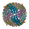
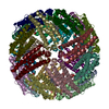
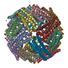
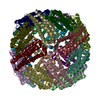
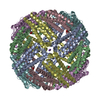
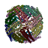
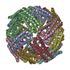
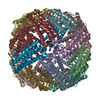
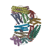
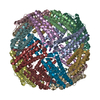
- PDB-2vxi: The binding of heme and zinc in Escherichia coli Bacterioferritin -
+ Open data

- Basic information
| Entry | Database: PDB / ID: 2vxi | ||||||
|---|---|---|---|---|---|---|---|
| Title | The binding of heme and zinc in Escherichia coli Bacterioferritin | ||||||
 Components Components | BACTERIOFERRITIN | ||||||
 Keywords Keywords | METAL TRANSPORT / BACTERIOFERRITIN / IRON STORAGE AND ELECTRON TRANSPORT / ZINC / HEME / IRON / IRON STORAGE / METAL-BINDING | ||||||
| Function / homology |  Function and homology information Function and homology informationiron ion sequestering activity / ferroxidase / ferroxidase activity / ferric iron binding / iron ion transport / intracellular iron ion homeostasis / oxidoreductase activity / iron ion binding / heme binding / protein homodimerization activity ...iron ion sequestering activity / ferroxidase / ferroxidase activity / ferric iron binding / iron ion transport / intracellular iron ion homeostasis / oxidoreductase activity / iron ion binding / heme binding / protein homodimerization activity / membrane / identical protein binding / cytosol Similarity search - Function | ||||||
| Biological species |  | ||||||
| Method |  X-RAY DIFFRACTION / SYNCHROTRON / MAD / Resolution: 1.91 Å X-RAY DIFFRACTION / SYNCHROTRON / MAD / Resolution: 1.91 Å | ||||||
 Authors Authors | Willies, S.C. / Isupov, M.N. / Garman, E.F. / Littlechild, J.A. | ||||||
 Citation Citation | Journal: J.Biol.Inorg.Chem. / Year: 2009 Title: The Binding of Haem and Zinc in the 1.9 A X-Ray Structure of Escherichia Coli Bacterioferritin. Authors: Willies, S.C. / Isupov, M.N. / Garman, E.F. / Littlechild, J.A. | ||||||
| History |
|
- Structure visualization
| Structure viewer | Molecule: MolmilJmol/JSmol |
|---|
- Downloads & links
-Download
| PDBx/mmCIF format | 2vxi.cif.gz | 457.8 KB | Display | PDBx/mmCIF format |
|---|---|---|---|---|
| PDB format | pdb2vxi.ent.gz | 384.9 KB | Display | PDB format |
| PDBx/mmJSON format | 2vxi.json.gz | Tree view | PDBx/mmJSON format | |
| Others |  Other downloads Other downloads |
-Validation report
| Arichive directory | https://data.pdbj.org/pub/pdb/validation_reports/vx/2vxiftp://data.pdbj.org/pub/pdb/validation_reports/vx/2vxi | HTTPS FTP |
|---|
-Related structure data
| Related structure data | |
|---|---|
| Similar structure data |


-Links
 PDBj
PDBj


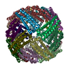
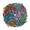
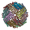
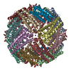
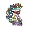
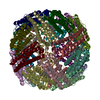
- Assembly
| Deposited unit | 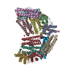
| ||||||||||||||||||||||||||||||||||||||||||||||||
|---|---|---|---|---|---|---|---|---|---|---|---|---|---|---|---|---|---|---|---|---|---|---|---|---|---|---|---|---|---|---|---|---|---|---|---|---|---|---|---|---|---|---|---|---|---|---|---|---|---|
| 1 | 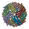
| ||||||||||||||||||||||||||||||||||||||||||||||||
| Unit cell |
| ||||||||||||||||||||||||||||||||||||||||||||||||
| Components on special symmetry positions |
| ||||||||||||||||||||||||||||||||||||||||||||||||
| Noncrystallographic symmetry (NCS) | NCS oper:
|
-Components
| #1: Protein | Mass: 18518.016 Da / Num. of mol.: 12 / Source method: isolated from a natural source / Source: (natural) #2: Chemical | ChemComp-HEM /   Mass: 616.487 Da / Num. of mol.: 12 / Source method: obtained synthetically / Formula: C34H32FeN4O4 Mass: 616.487 Da / Num. of mol.: 12 / Source method: obtained synthetically / Formula: C34H32FeN4O4#3: Chemical | ChemComp-ZN /   Mass: 65.409 Da / Num. of mol.: 24 / Source method: obtained synthetically / Formula: Zn Mass: 65.409 Da / Num. of mol.: 24 / Source method: obtained synthetically / Formula: Zn#4: Chemical | ChemComp-SO4 /   Mass: 96.063 Da / Num. of mol.: 29 / Source method: obtained synthetically / Formula: SO4 Mass: 96.063 Da / Num. of mol.: 29 / Source method: obtained synthetically / Formula: SO4#5: Water | ChemComp-HOH / |  Mass: 18.015 Da / Num. of mol.: 2208 / Source method: isolated from a natural source / Formula: H2O Mass: 18.015 Da / Num. of mol.: 2208 / Source method: isolated from a natural source / Formula: H2O |
|---|
-Experimental details
-Experiment
| Experiment | Method: X-RAY DIFFRACTION / Number of used crystals: 1 |
|---|
- Sample preparation
| Crystal | Density Matthews: 2.93 Å3/Da / Density % sol: 57.74 % / Description: NONE |
|---|---|
| Crystal grow | pH: 7.5 / Details: 60% AMSO4, 20MM TRIS-HCL PH7.5, 0.1M NACL |
-Data collection
| Diffraction | Mean temperature: 100 K | |||||||||
|---|---|---|---|---|---|---|---|---|---|---|
| Diffraction source | Source: SYNCHROTRON / Site: SRS  / Beamline: PX10.1 / Wavelength: 1.739, 1.729 / Beamline: PX10.1 / Wavelength: 1.739, 1.729 | |||||||||
| Detector | Type: MARRESEARCH / Detector: CCD / Date: Nov 17, 2005 | |||||||||
| Radiation | Protocol: MAD / Monochromatic (M) / Laue (L): M / Scattering type: x-ray | |||||||||
| Radiation wavelength |
| |||||||||
| Reflection | Resolution: 1.9→25 Å / Num. obs: 460546 / % possible obs: 97.7 % / Observed criterion σ(I): 0 / Redundancy: 3.77 % / Rmerge(I) obs: 0.1 / Net I/σ(I): 11 | |||||||||
| Reflection shell | Resolution: 1.9→1.93 Å / Redundancy: 2.2 % / Rmerge(I) obs: 0.38 / Mean I/σ(I) obs: 2.14 / % possible all: 90.2 |
- Processing
| Software |
| ||||||||||||||||||||||||||||||||||||||||||||||||||||||||||||||||||||||||||||||||||||||||||||||||||||||||||||||||||||||||||||||||||||||||||||||||||||||||||||||||||||||||||||||||||||||
|---|---|---|---|---|---|---|---|---|---|---|---|---|---|---|---|---|---|---|---|---|---|---|---|---|---|---|---|---|---|---|---|---|---|---|---|---|---|---|---|---|---|---|---|---|---|---|---|---|---|---|---|---|---|---|---|---|---|---|---|---|---|---|---|---|---|---|---|---|---|---|---|---|---|---|---|---|---|---|---|---|---|---|---|---|---|---|---|---|---|---|---|---|---|---|---|---|---|---|---|---|---|---|---|---|---|---|---|---|---|---|---|---|---|---|---|---|---|---|---|---|---|---|---|---|---|---|---|---|---|---|---|---|---|---|---|---|---|---|---|---|---|---|---|---|---|---|---|---|---|---|---|---|---|---|---|---|---|---|---|---|---|---|---|---|---|---|---|---|---|---|---|---|---|---|---|---|---|---|---|---|---|---|---|
| Refinement | Method to determine structure: MAD Starting model: NONE Resolution: 1.91→147.44 Å / Cor.coef. Fo:Fc: 0.976 / Cor.coef. Fo:Fc free: 0.966 / Cross valid method: THROUGHOUT / ESU R: 0.12 / ESU R Free: 0.119 / Stereochemistry target values: MAXIMUM LIKELIHOOD / Details: HYDROGENS HAVE BEEN ADDED IN THE RIDING POSITIONS.
| ||||||||||||||||||||||||||||||||||||||||||||||||||||||||||||||||||||||||||||||||||||||||||||||||||||||||||||||||||||||||||||||||||||||||||||||||||||||||||||||||||||||||||||||||||||||
| Solvent computation | Ion probe radii: 0.8 Å / Shrinkage radii: 0.8 Å / VDW probe radii: 1.2 Å / Solvent model: BABINET MODEL WITH MASK | ||||||||||||||||||||||||||||||||||||||||||||||||||||||||||||||||||||||||||||||||||||||||||||||||||||||||||||||||||||||||||||||||||||||||||||||||||||||||||||||||||||||||||||||||||||||
| Displacement parameters | Biso mean: 24.852 Å2
| ||||||||||||||||||||||||||||||||||||||||||||||||||||||||||||||||||||||||||||||||||||||||||||||||||||||||||||||||||||||||||||||||||||||||||||||||||||||||||||||||||||||||||||||||||||||
| Refinement step | Cycle: LAST / Resolution: 1.91→147.44 Å
| ||||||||||||||||||||||||||||||||||||||||||||||||||||||||||||||||||||||||||||||||||||||||||||||||||||||||||||||||||||||||||||||||||||||||||||||||||||||||||||||||||||||||||||||||||||||
| Refine LS restraints |
|