+ データを開く
- 基本情報
基本情報
| 登録情報 | データベース: PDB / ID: 6p4l | ||||||||||||||||||||||||||||||||||||||||||
|---|---|---|---|---|---|---|---|---|---|---|---|---|---|---|---|---|---|---|---|---|---|---|---|---|---|---|---|---|---|---|---|---|---|---|---|---|---|---|---|---|---|---|---|
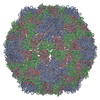
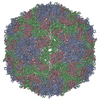
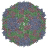
| タイトル | Bile salts alter the mouse norovirus capsid conformation; possible implications for cell attachment and immune evasion. | ||||||||||||||||||||||||||||||||||||||||||
 要素 要素 | Capsid protein | ||||||||||||||||||||||||||||||||||||||||||
 キーワード キーワード | VIRUS / murine / norovirus / VIRAL PROTEIN | ||||||||||||||||||||||||||||||||||||||||||
| 機能・相同性 |  機能・相同性情報 機能・相同性情報antigenic variation / symbiont-mediated perturbation of host defense response / virion component / host cell cytoplasm 類似検索 - 分子機能 | ||||||||||||||||||||||||||||||||||||||||||
| 生物種 |   Murine norovirus 1 (マウスノロウイルス 1) Murine norovirus 1 (マウスノロウイルス 1) | ||||||||||||||||||||||||||||||||||||||||||
| 手法 | 電子顕微鏡法 / 単粒子再構成法 / クライオ電子顕微鏡法 / 解像度: 3.1 Å | ||||||||||||||||||||||||||||||||||||||||||
 データ登録者 データ登録者 | Smith, T.J. | ||||||||||||||||||||||||||||||||||||||||||
| 資金援助 |  米国, 1件 米国, 1件
| ||||||||||||||||||||||||||||||||||||||||||
 引用 引用 |  ジャーナル: J Virol / 年: 2019 ジャーナル: J Virol / 年: 2019タイトル: Bile Salts Alter the Mouse Norovirus Capsid Conformation: Possible Implications for Cell Attachment and Immune Evasion. 著者: Michael B Sherman / Alexis N Williams / Hong Q Smith / Christopher Nelson / Craig B Wilen / Daved H Fremont / Herbert W Virgin / Thomas J Smith /  要旨: Caliciviruses are single-stranded RNA viruses with 180 copies of capsid protein comprising the T=3 icosahedral capsids. The main capsid feature is a pronounced protruding (P) domain dimer formed by ...Caliciviruses are single-stranded RNA viruses with 180 copies of capsid protein comprising the T=3 icosahedral capsids. The main capsid feature is a pronounced protruding (P) domain dimer formed by adjacent subunits on the icosahedral surface while the shell domain forms a tight icosahedral sphere around the genome. While the P domain in the crystal structure of human Norwalk virus (genotype I.1) was tightly associated with the shell surface, the cryo-electron microscopy (cryo-EM) structures of several members of the family (mouse norovirus [MNV], rabbit hemorrhagic disease virus, and human norovirus genotype II.10) revealed a "floating" P domain that hovers above the shell by nearly 10 to 15 Å in physiological buffers. Since this unusual feature is shared among, and unique to, the , it suggests an important biological role. Recently, we demonstrated that bile salts enhance cell attachment to the target cell and increase the intrinsic affinity between the P domain and receptor. Presented here are the cryo-EM structures of MNV-1 in the presence of bile salts (∼3 Å) and the receptor CD300lf (∼8 Å). Surprisingly, bile salts cause the rotation and contraction of the P domain onto the shell surface. This both stabilizes the P domain and appears to allow for a higher degree of saturation of receptor onto the virus. Together, these results suggest that, as the virus moves into the gut and the associated high concentrations of bile, the entire capsid face undergoes a conformational change to optimize receptor avidity while the P domain itself undergoes smaller conformational changes to improve receptor affinity. Mouse norovirus and several other members of the have been shown to have a highly unusual structure with the receptor binding protruding (P) domain only loosely tethered to the main capsid shell. Recent studies demonstrated that bile salts enhance the intrinsic P domain/receptor affinity and is necessary for cell attachment. Presented here are the high-resolution cryo-EM structures of apo MNV, MNV/bile salt, and MNV/bile salt/receptor. Bile salts cause a 90° rotation and collapse of the P domain onto the shell surface that may increase the number of available receptor binding sites. Therefore, bile salts appear to be having several effects on MNV. Bile salts shift the structural equilibrium of the P domain toward a form that binds the receptor and away from one that binds antibody. They may also cause the entire P domain to optimize receptor binding while burying a number of potential epitopes. | ||||||||||||||||||||||||||||||||||||||||||
| 履歴 |
|
- 構造の表示
構造の表示
| ムービー |
 ムービービューア ムービービューア |
|---|---|
| 構造ビューア | 分子:  Molmil Molmil Jmol/JSmol Jmol/JSmol |
- ダウンロードとリンク
- ダウンロード
| PDBx/mmCIF形式 |  6p4l.cif.gz 6p4l.cif.gz | 128 KB | 表示 |  PDBx/mmCIF形式 PDBx/mmCIF形式 |
|---|---|---|---|---|
| PDB形式 |  pdb6p4l.ent.gz pdb6p4l.ent.gz | 91.7 KB | 表示 |  PDB形式 PDB形式 |
| PDBx/mmJSON形式 |  6p4l.json.gz 6p4l.json.gz | ツリー表示 |  PDBx/mmJSON形式 PDBx/mmJSON形式 | |
| その他 |  その他のダウンロード その他のダウンロード |
-検証レポート
| 文書・要旨 |  6p4l_validation.pdf.gz 6p4l_validation.pdf.gz | 849.1 KB | 表示 |  wwPDB検証レポート wwPDB検証レポート |
|---|---|---|---|---|
| 文書・詳細版 |  6p4l_full_validation.pdf.gz 6p4l_full_validation.pdf.gz | 855.8 KB | 表示 | |
| XML形式データ |  6p4l_validation.xml.gz 6p4l_validation.xml.gz | 25.6 KB | 表示 | |
| CIF形式データ |  6p4l_validation.cif.gz 6p4l_validation.cif.gz | 36.8 KB | 表示 | |
| アーカイブディレクトリ |  https://data.pdbj.org/pub/pdb/validation_reports/p4/6p4l https://data.pdbj.org/pub/pdb/validation_reports/p4/6p4l ftp://data.pdbj.org/pub/pdb/validation_reports/p4/6p4l ftp://data.pdbj.org/pub/pdb/validation_reports/p4/6p4l | HTTPS FTP |
-関連構造データ
- リンク
リンク
- 集合体
集合体
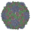
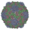
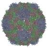
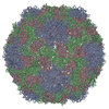
| 登録構造単位 | 
|
|---|---|
| 1 | x 60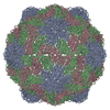
|
| 2 |
|
| 3 | x 5
|
| 4 | x 6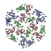
|
| 5 | 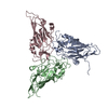
|
| 対称性 | 点対称性: (シェーンフリース記号: I (正20面体型対称)) |
- 要素
要素
| #1: タンパク質 | 分子量: 58700.598 Da / 分子数: 3 / 由来タイプ: 組換発現 由来: (組換発現)   Murine norovirus 1 (マウスノロウイルス 1) Murine norovirus 1 (マウスノロウイルス 1)発現宿主:   Murine norovirus 1 (マウスノロウイルス 1) Murine norovirus 1 (マウスノロウイルス 1)参照: UniProt: Q2V8W4 Has protein modification | N | |
|---|
-実験情報
-実験
| 実験 | 手法: 電子顕微鏡法 |
|---|---|
| EM実験 | 試料の集合状態: PARTICLE / 3次元再構成法: 単粒子再構成法 |
- 試料調製
試料調製
| 構成要素 | 名称: Murine norovirus 1 / タイプ: VIRUS / Entity ID: all / 由来: NATURAL |
|---|---|
| 由来(天然) | 生物種:   Murine norovirus 1 (マウスノロウイルス 1) Murine norovirus 1 (マウスノロウイルス 1) |
| ウイルスについての詳細 | 中空か: NO / エンベロープを持つか: NO / 単離: STRAIN / タイプ: VIRION |
| 緩衝液 | pH: 7.2 |
| 試料 | 包埋: NO / シャドウイング: NO / 染色: NO / 凍結: YES |
| 急速凍結 | 凍結剤: ETHANE |
- 電子顕微鏡撮影
| 実験機器 |  モデル: Titan Krios / 画像提供: FEI Company |
|---|---|
| 顕微鏡 | モデル: FEI TITAN KRIOS |
| 電子銃 | 電子線源:  FIELD EMISSION GUN / 加速電圧: 300 kV / 照射モード: SPOT SCAN FIELD EMISSION GUN / 加速電圧: 300 kV / 照射モード: SPOT SCAN |
| 電子レンズ | モード: BRIGHT FIELD |
| 撮影 | 電子線照射量: 28 e/Å2 / フィルム・検出器のモデル: OTHER |
- 解析
解析
| ソフトウェア | 名称: PHENIX / バージョン: 1.15_3448: / 分類: 精密化 | ||||||||||||||||||||||||
|---|---|---|---|---|---|---|---|---|---|---|---|---|---|---|---|---|---|---|---|---|---|---|---|---|---|
| EMソフトウェア | 名称: PHENIX / カテゴリ: モデル精密化 | ||||||||||||||||||||||||
| CTF補正 | タイプ: PHASE FLIPPING AND AMPLITUDE CORRECTION | ||||||||||||||||||||||||
| 3次元再構成 | 解像度: 3.1 Å / 解像度の算出法: FSC 0.143 CUT-OFF / 粒子像の数: 30719 / 対称性のタイプ: POINT | ||||||||||||||||||||||||
| 拘束条件 |
|
 ムービー
ムービー コントローラー




 PDBj
PDBj
