 Movie
Movie Controller

[English] 日本語
 Yorodumi
- PDB-6p2h: Structural basis for 2'-deoxyguanosine recognition by the 2'-dG-I... -
+ Open data

- Basic information
| Entry | Database: PDB / ID: 6p2h | ||||||
|---|---|---|---|---|---|---|---|
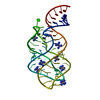
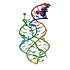
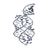
| Title | Structural basis for 2'-deoxyguanosine recognition by the 2'-dG-II class of riboswitches | ||||||
 Components Components | RNA (69-MER) | ||||||
 Keywords Keywords | RNA / riboswitch aptamer 2'-deoxyguanosine purine nucleoside | ||||||
| Function / homology | 2'-DEOXY-GUANOSINE / COBALT HEXAMMINE(III) / RNA / RNA (> 10) Function and homology information Function and homology information | ||||||
| Biological species | synthetic construct (others) | ||||||
| Method |  X-RAY DIFFRACTION / MOLECULAR REPLACEMENT / Resolution: 2.803 Å X-RAY DIFFRACTION / MOLECULAR REPLACEMENT / Resolution: 2.803 Å | ||||||
 Authors Authors | Matyjasik, M.M. / Batey, R.T. | ||||||
| Funding support |  United States, 1items United States, 1items
| ||||||
 Citation Citation | Journal: Nucleic Acids Res. / Year: 2019 Title: Structural basis for 2'-deoxyguanosine recognition by the 2'-dG-II class of riboswitches. Authors: Matyjasik, M.M. / Batey, R.T. | ||||||
| History |
|
- Structure visualization
| Structure viewer | Molecule: MolmilJmol/JSmol |
|---|
- Downloads & links
-Download
| PDBx/mmCIF format | 6p2h.cif.gz | 54.1 KB | Display | PDBx/mmCIF format |
|---|---|---|---|---|
| PDB format | pdb6p2h.ent.gz | 36.6 KB | Display | PDB format |
| PDBx/mmJSON format | 6p2h.json.gz | Tree view | PDBx/mmJSON format | |
| Others |  Other downloads Other downloads |
-Validation report
| Arichive directory | https://data.pdbj.org/pub/pdb/validation_reports/p2/6p2hftp://data.pdbj.org/pub/pdb/validation_reports/p2/6p2h | HTTPS FTP |
|---|
-Related structure data
| Related structure data | 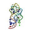 4fe5S S: Starting model for refinement |
|---|---|
| Similar structure data |


-Links
 PDBj
PDBj


- Assembly
| Deposited unit | 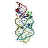
| ||||||||||
|---|---|---|---|---|---|---|---|---|---|---|---|
| 1 |
| ||||||||||
| Unit cell |
| ||||||||||
| Components on special symmetry positions |
|
-Components
| #1: RNA chain | Mass: 22323.186 Da / Num. of mol.: 1 / Source method: obtained synthetically / Source: (synth.) synthetic construct (others) | ||||
|---|---|---|---|---|---|
| #2: Chemical | ChemComp-GNG /   Mass: 267.241 Da / Num. of mol.: 1 / Source method: obtained synthetically / Formula: C10H13N5O4 / Feature type: SUBJECT OF INVESTIGATION Mass: 267.241 Da / Num. of mol.: 1 / Source method: obtained synthetically / Formula: C10H13N5O4 / Feature type: SUBJECT OF INVESTIGATION | ||||
| #3: Chemical | ChemComp-NCO /   Mass: 161.116 Da / Num. of mol.: 5 / Source method: obtained synthetically / Formula: CoH18N6 Mass: 161.116 Da / Num. of mol.: 5 / Source method: obtained synthetically / Formula: CoH18N6#4: Chemical | ChemComp-MG /   Mass: 24.305 Da / Num. of mol.: 5 / Source method: obtained synthetically / Formula: Mg Mass: 24.305 Da / Num. of mol.: 5 / Source method: obtained synthetically / Formula: Mg#5: Water | ChemComp-HOH / |  Mass: 18.015 Da / Num. of mol.: 12 / Source method: isolated from a natural source / Formula: H2O Mass: 18.015 Da / Num. of mol.: 12 / Source method: isolated from a natural source / Formula: H2O |
-Experimental details
-Experiment
| Experiment | Method: X-RAY DIFFRACTION / Number of used crystals: 1 |
|---|
- Sample preparation
| Crystal | Density Matthews: 3.67 Å3/Da / Density % sol: 66.48 % / Description: Tetragonal |
|---|---|
| Crystal grow | Temperature: 303 K / Method: vapor diffusion, hanging drop / pH: 6 Details: 40 mM sodium cacodylate pH 6.0, 23% v/v methylpentanediol, 18 mM cobalt hexamine, 80 mM potassium chloride, and 12 mM sodium chloride |
-Data collection
| Diffraction | Mean temperature: 100 K / Ambient temp details: Liquid nitrogen cryostream / Serial crystal experiment: N |
|---|---|
| Diffraction source | Source: ROTATING ANODE / Type: RIGAKU MICROMAX-007 HF / Wavelength: 1.5406 Å |
| Detector | Type: DECTRIS PILATUS 200K / Detector: PIXEL / Date: Jun 22, 2018 |
| Radiation | Monochromator: Cu / Protocol: SINGLE WAVELENGTH / Monochromatic (M) / Laue (L): M / Scattering type: x-ray |
| Radiation wavelength | Wavelength: 1.5406 Å / Relative weight: 1 |
| Reflection | Resolution: 2.803→29.953 Å / Num. obs: 8547 / % possible obs: 97.69 % / Redundancy: 10.8 % / Biso Wilson estimate: 58.19 Å2 / Rmerge(I) obs: 0.096 / Rpim(I) all: 0.03 / Χ2: 1.99 / Net I/σ(I): 27.25 |
| Reflection shell | Resolution: 2.803→2.904 Å / Redundancy: 10.3 % / Rmerge(I) obs: 0.608 / Mean I/σ(I) obs: 3.37 / Num. unique obs: 816 / CC1/2: 0.946 / Rpim(I) all: 0.195 / Χ2: 1.515 / % possible all: 97.69 |
- Processing
| Software |
| |||||||||||||||||||||||||||||||||||||||||||||||||
|---|---|---|---|---|---|---|---|---|---|---|---|---|---|---|---|---|---|---|---|---|---|---|---|---|---|---|---|---|---|---|---|---|---|---|---|---|---|---|---|---|---|---|---|---|---|---|---|---|---|---|
| Refinement | Method to determine structure: MOLECULAR REPLACEMENT Starting model: 4FE5 Resolution: 2.803→29.953 Å / SU ML: 0.47 / Cross valid method: FREE R-VALUE / σ(F): 1.33 / Phase error: 30
| |||||||||||||||||||||||||||||||||||||||||||||||||
| Solvent computation | Shrinkage radii: 0.9 Å / VDW probe radii: 1.11 Å | |||||||||||||||||||||||||||||||||||||||||||||||||
| Refinement step | Cycle: LAST / Resolution: 2.803→29.953 Å
| |||||||||||||||||||||||||||||||||||||||||||||||||
| Refine LS restraints |
| |||||||||||||||||||||||||||||||||||||||||||||||||
| LS refinement shell |
|
