 Movie
Movie Controller


+ Open data

- Basic information
| Entry | Database: PDB / ID: 6p24 | ||||||
|---|---|---|---|---|---|---|---|
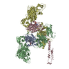
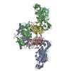
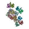
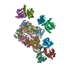
| Title | Escherichia coli tRNA synthetase | ||||||
 Components Components | (Phenylalanine--tRNA ligase ...) x 2 | ||||||
 Keywords Keywords | LIGASE / Inhibitor / Aminoacyl-tRNA synthetase / PheRS / Antibacterial | ||||||
| Function / homology |  Function and homology information Function and homology informationphenylalanine-tRNA ligase complex / phenylalanine-tRNA ligase / phenylalanyl-tRNA aminoacylation / phenylalanine-tRNA ligase activity / tRNA binding / magnesium ion binding / ATP binding / membrane / identical protein binding / cytoplasm / cytosol Similarity search - Function | ||||||
| Biological species |  | ||||||
| Method |  X-RAY DIFFRACTION / SYNCHROTRON / MOLECULAR REPLACEMENT / Resolution: 2.12 Å X-RAY DIFFRACTION / SYNCHROTRON / MOLECULAR REPLACEMENT / Resolution: 2.12 Å | ||||||
 Authors Authors | Kahne, D. / Baidin, V. / Owens, T.W. | ||||||
| Funding support |  United States, 1items United States, 1items
| ||||||
 Citation Citation | Journal: J.Am.Chem.Soc. / Year: 2021 Title: Simple Secondary Amines Inhibit Growth of Gram-Negative Bacteria through Highly Selective Binding to Phenylalanyl-tRNA Synthetase. Authors: Baidin, V. / Owens, T.W. / Lazarus, M.B. / Kahne, D. | ||||||
| History |
|
- Structure visualization
| Structure viewer | Molecule: MolmilJmol/JSmol |
|---|
- Downloads & links
-Download
| PDBx/mmCIF format | 6p24.cif.gz | 549.2 KB | Display | PDBx/mmCIF format |
|---|---|---|---|---|
| PDB format | pdb6p24.ent.gz | 354.4 KB | Display | PDB format |
| PDBx/mmJSON format | 6p24.json.gz | Tree view | PDBx/mmJSON format | |
| Others |  Other downloads Other downloads |
-Validation report
| Arichive directory | https://data.pdbj.org/pub/pdb/validation_reports/p2/6p24ftp://data.pdbj.org/pub/pdb/validation_reports/p2/6p24 | HTTPS FTP |
|---|
-Related structure data
| Related structure data |  6oz5C 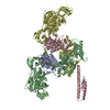 6p26C 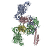 6p8tC  3pcoS S: Starting model for refinement C: citing same article ( |
|---|---|
| Similar structure data |


-Links
 PDBj
PDBj

- Assembly
| Deposited unit | 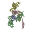
| ||||||||||
|---|---|---|---|---|---|---|---|---|---|---|---|
| 1 |
| ||||||||||
| Unit cell |
|
-Components
-Phenylalanine--tRNA ligase ... , 2 types, 4 molecules ACBD
| #1: Protein | Mass: 37318.156 Da / Num. of mol.: 2 Source method: isolated from a genetically manipulated source Source: (gene. exp.) Strain: K12 / Gene: pheS, b1714, JW5277 / Production host: #2: Protein | Mass: 87483.039 Da / Num. of mol.: 2 Source method: isolated from a genetically manipulated source Source: (gene. exp.) Strain: K12 / Gene: pheT, b1713, JW1703 / Production host: |
|---|
-Non-polymers , 8 types, 579 molecules 


| #3: Chemical | ChemComp-MG /  Mass: 24.305 Da / Num. of mol.: 13 / Source method: obtained synthetically / Formula: Mg Mass: 24.305 Da / Num. of mol.: 13 / Source method: obtained synthetically / Formula: Mg#4: Chemical | ChemComp-CL / |  Mass: 35.453 Da / Num. of mol.: 1 / Source method: obtained synthetically / Formula: Cl Mass: 35.453 Da / Num. of mol.: 1 / Source method: obtained synthetically / Formula: Cl#5: Chemical | ChemComp-HEZ /  Mass: 118.174 Da / Num. of mol.: 5 / Source method: obtained synthetically / Formula: C6H14O2 Mass: 118.174 Da / Num. of mol.: 5 / Source method: obtained synthetically / Formula: C6H14O2#6: Chemical | ChemComp-TRS /  Mass: 122.143 Da / Num. of mol.: 4 / Source method: obtained synthetically / Formula: C4H12NO3 / Comment: pH buffer*YM Mass: 122.143 Da / Num. of mol.: 4 / Source method: obtained synthetically / Formula: C4H12NO3 / Comment: pH buffer*YM#7: Chemical | ChemComp-PEG /  Mass: 106.120 Da / Num. of mol.: 10 / Source method: obtained synthetically / Formula: C4H10O3 Mass: 106.120 Da / Num. of mol.: 10 / Source method: obtained synthetically / Formula: C4H10O3#8: Chemical | ChemComp-EDO /  Mass: 62.068 Da / Num. of mol.: 17 / Source method: obtained synthetically / Formula: C2H6O2 Mass: 62.068 Da / Num. of mol.: 17 / Source method: obtained synthetically / Formula: C2H6O2#9: Chemical |  Mass: 150.173 Da / Num. of mol.: 2 / Source method: obtained synthetically / Formula: C6H14O4 Mass: 150.173 Da / Num. of mol.: 2 / Source method: obtained synthetically / Formula: C6H14O4#10: Water | ChemComp-HOH / | Mass: 18.015 Da / Num. of mol.: 527 / Source method: isolated from a natural source / Formula: H2O |
|---|
-Details
| Has ligand of interest | N |
|---|
-Experimental details
-Experiment
| Experiment | Method: X-RAY DIFFRACTION / Number of used crystals: 1 |
|---|
- Sample preparation
| Crystal | Density Matthews: 2.68 Å3/Da / Density % sol: 54.08 % |
|---|---|
| Crystal grow | Temperature: 293 K / Method: vapor diffusion, hanging drop / pH: 8.5 Details: 16% PEG 8,000, 200 mM MgCl2, 100 mM Tris-HCl pH8.5, 2% 1,6-hexanediol, 3% d-Galactose |
-Data collection
| Diffraction | Mean temperature: 100 K / Serial crystal experiment: N |
|---|---|
| Diffraction source | Source: SYNCHROTRON / Site: APS / Beamline: 24-ID-E / Wavelength: 0.98 Å |
| Detector | Type: DECTRIS EIGER X 16M / Detector: PIXEL / Date: Dec 14, 2017 |
| Radiation | Protocol: SINGLE WAVELENGTH / Monochromatic (M) / Laue (L): M / Scattering type: x-ray |
| Radiation wavelength | Wavelength: 0.98 Å / Relative weight: 1 |
| Reflection | Resolution: 2.115→173.58 Å / Num. obs: 152649 / % possible obs: 98.7 % / Redundancy: 3.3 % / Biso Wilson estimate: 50.86 Å2 / CC1/2: 0.994 / Net I/σ(I): 6.7 |
| Reflection shell | Resolution: 2.115→2.15 Å / Num. unique obs: 6642 / CC1/2: 0.126 |
- Processing
| Software |
| |||||||||||||||||||||||||||||||||||||||||||||||||||||||||||||||||||||||||||||||||||||||||||||||||||||||||
|---|---|---|---|---|---|---|---|---|---|---|---|---|---|---|---|---|---|---|---|---|---|---|---|---|---|---|---|---|---|---|---|---|---|---|---|---|---|---|---|---|---|---|---|---|---|---|---|---|---|---|---|---|---|---|---|---|---|---|---|---|---|---|---|---|---|---|---|---|---|---|---|---|---|---|---|---|---|---|---|---|---|---|---|---|---|---|---|---|---|---|---|---|---|---|---|---|---|---|---|---|---|---|---|---|---|---|
| Refinement | Method to determine structure: MOLECULAR REPLACEMENT Starting model: 3PCO Resolution: 2.12→82.04 Å / SU ML: 0.351 / Cross valid method: FREE R-VALUE / σ(F): 1.33 / Phase error: 28.6791 Stereochemistry target values: GeoStd + Monomer Library + CDL v1.2
| |||||||||||||||||||||||||||||||||||||||||||||||||||||||||||||||||||||||||||||||||||||||||||||||||||||||||
| Solvent computation | Shrinkage radii: 0.9 Å / VDW probe radii: 1.11 Å / Solvent model: FLAT BULK SOLVENT MODEL | |||||||||||||||||||||||||||||||||||||||||||||||||||||||||||||||||||||||||||||||||||||||||||||||||||||||||
| Displacement parameters | Biso mean: 60.39 Å2 | |||||||||||||||||||||||||||||||||||||||||||||||||||||||||||||||||||||||||||||||||||||||||||||||||||||||||
| Refinement step | Cycle: LAST / Resolution: 2.12→82.04 Å
| |||||||||||||||||||||||||||||||||||||||||||||||||||||||||||||||||||||||||||||||||||||||||||||||||||||||||
| Refine LS restraints |
| |||||||||||||||||||||||||||||||||||||||||||||||||||||||||||||||||||||||||||||||||||||||||||||||||||||||||
| LS refinement shell |
|
