 Movie
Movie Controller

[English] 日本語
 Yorodumi
- PDB-6ov3: Crystal structure of human claudin-9 in complex with Clostridium ... -
+ Open data

- Basic information
| Entry | Database: PDB / ID: 6ov3 | |||||||||
|---|---|---|---|---|---|---|---|---|---|---|
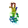
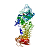
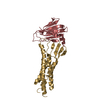
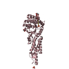
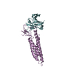
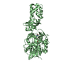
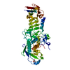
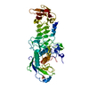
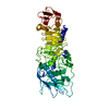
| Title | Crystal structure of human claudin-9 in complex with Clostridium perfringens entertoxin C-terminal domain in open form | |||||||||
 Components Components |
| |||||||||
 Keywords Keywords | CELL ADHESION / Claudin / Enterotoxin / Tight junction protein / Transmembrane protein | |||||||||
| Function / homology |  Function and homology information Function and homology informationcalcium-independent cell-cell adhesion / Tight junction interactions / bicellular tight junction assembly / bicellular tight junction / cell junction / toxin activity / virus receptor activity / cell adhesion / structural molecule activity / extracellular region ...calcium-independent cell-cell adhesion / Tight junction interactions / bicellular tight junction assembly / bicellular tight junction / cell junction / toxin activity / virus receptor activity / cell adhesion / structural molecule activity / extracellular region / identical protein binding / plasma membrane Similarity search - Function | |||||||||
| Biological species |  Homo sapiens (human) Homo sapiens (human)  Clostridium perfringens (bacteria) Clostridium perfringens (bacteria) | |||||||||
| Method |  X-RAY DIFFRACTION / SYNCHROTRON / MOLECULAR REPLACEMENT / Resolution: 3.25 Å X-RAY DIFFRACTION / SYNCHROTRON / MOLECULAR REPLACEMENT / Resolution: 3.25 Å | |||||||||
 Authors Authors | Vecchio, A.J. / Stroud, R.M. | |||||||||
| Funding support |  United States, 2items United States, 2items
| |||||||||
 Citation Citation | Journal: Proc.Natl.Acad.Sci.USA / Year: 2019 Title: Claudin-9 structures reveal mechanism for toxin-induced gut barrier breakdown. Authors: Vecchio, A.J. / Stroud, R.M. | |||||||||
| History |
|
- Structure visualization
| Structure viewer | Molecule: MolmilJmol/JSmol |
|---|
- Downloads & links
-Download
| PDBx/mmCIF format | 6ov3.cif.gz | 72 KB | Display | PDBx/mmCIF format |
|---|---|---|---|---|
| PDB format | pdb6ov3.ent.gz | 51.6 KB | Display | PDB format |
| PDBx/mmJSON format | 6ov3.json.gz | Tree view | PDBx/mmJSON format | |
| Others |  Other downloads Other downloads |
-Validation report
| Arichive directory | https://data.pdbj.org/pub/pdb/validation_reports/ov/6ov3ftp://data.pdbj.org/pub/pdb/validation_reports/ov/6ov3 | HTTPS FTP |
|---|
-Related structure data
| Related structure data |  6ov2C  3am2S C: citing same article ( S: Starting model for refinement |
|---|---|
| Similar structure data |
-Links
 PDBj
PDBj
- Assembly
| Deposited unit | 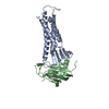
| ||||||||
|---|---|---|---|---|---|---|---|---|---|
| 1 |
| ||||||||
| Unit cell |
|
-Components
| #1: Protein | Mass: 22862.207 Da / Num. of mol.: 1 Source method: isolated from a genetically manipulated source Source: (gene. exp.) Homo sapiens (human) / Gene: CLDN9 / Cell line (production host): Tn5 / Production host:  Trichoplusia ni (cabbage looper) / References: UniProt: O95484 Trichoplusia ni (cabbage looper) / References: UniProt: O95484 |
|---|---|
| #2: Protein | Mass: 14795.565 Da / Num. of mol.: 1 / Fragment: C-terminal domain (UNP residues 194-319) Source method: isolated from a genetically manipulated source Source: (gene. exp.) Clostridium perfringens (bacteria) / Gene: cpe / Cell line (production host): Sf9 / Production host:  Spodoptera frugiperda (fall armyworm) / References: UniProt: P01558 Spodoptera frugiperda (fall armyworm) / References: UniProt: P01558 |
| Has protein modification | Y |
-Experimental details
-Experiment
| Experiment | Method: X-RAY DIFFRACTION / Number of used crystals: 1 |
|---|
- Sample preparation
| Crystal | Density Matthews: 6.12 Å3/Da / Density % sol: 79.92 % |
|---|---|
| Crystal grow | Temperature: 277 K / Method: vapor diffusion, sitting drop / pH: 4.5 Details: 150 mM sodium acetate, sodium cacodylate, Bis-Tris propane, pH 4.5, 25% PEG1500 |
-Data collection
| Diffraction | Mean temperature: 100 K / Serial crystal experiment: N |
|---|---|
| Diffraction source | Source: SYNCHROTRON / Site: ALS / Beamline: 8.3.1 / Wavelength: 1.11583 Å |
| Detector | Type: DECTRIS PILATUS3 S 6M / Detector: PIXEL / Date: Oct 20, 2017 |
| Radiation | Monochromator: double crystal Si(111) / Protocol: SINGLE WAVELENGTH / Monochromatic (M) / Laue (L): M / Scattering type: x-ray |
| Radiation wavelength | Wavelength: 1.11583 Å / Relative weight: 1 |
| Reflection | Resolution: 3.2→70.98 Å / Num. obs: 16155 / % possible obs: 100 % / Redundancy: 6.1 % / Biso Wilson estimate: 145.23 Å2 / CC1/2: 0.999 / Rmerge(I) obs: 0.127 / Rpim(I) all: 0.056 / Rrim(I) all: 0.139 / Net I/σ(I): 3.9 |
| Reflection shell | Resolution: 3.2→3.42 Å / Redundancy: 6.2 % / Rmerge(I) obs: 4.941 / Num. unique obs: 2877 / CC1/2: 0.512 / Rpim(I) all: 2.146 / Rrim(I) all: 5.397 / % possible all: 100 |
- Processing
| Software |
| ||||||||||||||||||||||||||||||||||||||||||||||||||||||||||||||||||||||||||||||||||||||||||||||||||||||||||||||||
|---|---|---|---|---|---|---|---|---|---|---|---|---|---|---|---|---|---|---|---|---|---|---|---|---|---|---|---|---|---|---|---|---|---|---|---|---|---|---|---|---|---|---|---|---|---|---|---|---|---|---|---|---|---|---|---|---|---|---|---|---|---|---|---|---|---|---|---|---|---|---|---|---|---|---|---|---|---|---|---|---|---|---|---|---|---|---|---|---|---|---|---|---|---|---|---|---|---|---|---|---|---|---|---|---|---|---|---|---|---|---|---|---|---|
| Refinement | Method to determine structure: MOLECULAR REPLACEMENT Starting model: Homology model and PDB entry 3AM2 Resolution: 3.25→20.125 Å / SU ML: 0.7 / Cross valid method: FREE R-VALUE / σ(F): 1.33 / Phase error: 44.59
| ||||||||||||||||||||||||||||||||||||||||||||||||||||||||||||||||||||||||||||||||||||||||||||||||||||||||||||||||
| Solvent computation | Shrinkage radii: 1.2 Å / VDW probe radii: 1.4 Å | ||||||||||||||||||||||||||||||||||||||||||||||||||||||||||||||||||||||||||||||||||||||||||||||||||||||||||||||||
| Displacement parameters | Biso mean: 185.27 Å2 | ||||||||||||||||||||||||||||||||||||||||||||||||||||||||||||||||||||||||||||||||||||||||||||||||||||||||||||||||
| Refinement step | Cycle: LAST / Resolution: 3.25→20.125 Å
| ||||||||||||||||||||||||||||||||||||||||||||||||||||||||||||||||||||||||||||||||||||||||||||||||||||||||||||||||
| Refine LS restraints |
| ||||||||||||||||||||||||||||||||||||||||||||||||||||||||||||||||||||||||||||||||||||||||||||||||||||||||||||||||
| LS refinement shell |
|
