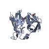
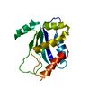
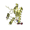
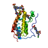
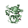
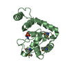
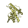
Entry Database : PDB / ID : 6otdTitle Globin sensor domain of AfGcHK in monomeric form, with imidazole Globin-coupled histidine kinase Keywords / / / / / / Function / homology Function Domain/homology Component
/ / / / / / / / / / / / / / / / / / / / / / / / / / / / / / / / Biological species Anaeromyxobacter sp. Fw109-5 (bacteria)Method / / / Resolution : 1.8 Å Authors Skalova, T. / Dohnalek, J. / Kolenko, P. / Stranava, M. / Lengalova, A. / Martinkova, M. Funding support Organization Grant number Country Ministry of Education (MoE, Czech Republic) LM2015043 European Regional Development Fund CZ.02.1.01/0.0/0.0/16_013/0001776
Journal : J.Biol.Chem. / Year : 2020Title : Disruption of the dimerization interface of the sensing domain in the dimeric heme-based oxygen sensorAfGcHK abolishes bacterial signal transduction.Authors : Skalova, T. / Lengalova, A. / Dohnalek, J. / Harlos, K. / Mihalcin, P. / Kolenko, P. / Stranava, M. / Blaha, J. / Shimizu, T. / Martinkova, M. History Deposition May 3, 2019 Deposition site / Processing site Revision 1.0 Jan 8, 2020 Provider / Type Revision 1.1 Jan 22, 2020 Group / Category / citation_authorItem / _citation.pdbx_database_id_PubMed / _citation.titleRevision 1.2 Feb 19, 2020 Group / Category / citation_authorItem _citation.journal_volume / _citation.page_first ... _citation.journal_volume / _citation.page_first / _citation.page_last / _citation.year / _citation_author.identifier_ORCID Revision 1.3 Oct 11, 2023 Group / Database references / Refinement descriptionCategory chem_comp_atom / chem_comp_bond ... chem_comp_atom / chem_comp_bond / database_2 / pdbx_initial_refinement_model Item / _database_2.pdbx_database_accession
Show all Show less
 Movie
Movie Controller


 Open data
 Basic information
 Components
 Keywords
 Function and homology information
 Anaeromyxobacter sp. Fw109-5 (bacteria)
 X-RAY DIFFRACTION /
  Authors
 Czech Republic, 2items
  Citation
 Structure visualization
 Downloads & links
 Other downloads
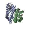


 PDBj
PDBj


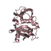
 Assembly
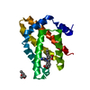

 Mass: 616.487 Da / Num. of mol.: 1 / Source method: obtained synthetically / Formula: C34H32FeN4O4
 Mass: 118.174 Da / Num. of mol.: 1 / Source method: obtained synthetically / Formula: C6H14O2 / Comment: precipitant*YM
 Mass: 69.085 Da / Num. of mol.: 2 / Source method: obtained synthetically / Formula: C3H5N2 / Feature type: SUBJECT OF INVESTIGATION
 Mass: 18.015 Da / Num. of mol.: 68 / Source method: isolated from a natural source / Formula: H2O
 Sample preparation
 / Beamline: I02 / Wavelength: 0.97 Å
 Processing