 Movie
Movie Controller

[English] 日本語
 Yorodumi
- PDB-6ojo: Dihydroneopterin aldolase (DHNA) from Yersinia pestis co-crystall... -
+ Open data

- Basic information
| Entry | Database: PDB / ID: 6ojo | ||||||
|---|---|---|---|---|---|---|---|
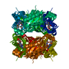
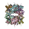
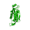
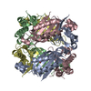
| Title | Dihydroneopterin aldolase (DHNA) from Yersinia pestis co-crystallized with pterine | ||||||
 Components Components | 7,8-dihydroneopterin aldolase | ||||||
 Keywords Keywords | BIOSYNTHETIC PROTEIN / folB folate pathway | ||||||
| Function / homology |  Function and homology information Function and homology informationdihydroneopterin aldolase / dihydroneopterin aldolase activity / folic acid biosynthetic process / tetrahydrofolate biosynthetic process / kinase activity / cytoplasm Similarity search - Function | ||||||
| Biological species |   Yersinia pestis (bacteria) Yersinia pestis (bacteria) | ||||||
| Method |  X-RAY DIFFRACTION / MOLECULAR REPLACEMENT / Resolution: 1.886 Å X-RAY DIFFRACTION / MOLECULAR REPLACEMENT / Resolution: 1.886 Å | ||||||
 Authors Authors | Bourne, C.R. | ||||||
| Funding support |  United States, 1items United States, 1items
| ||||||
 Citation Citation | Journal: To Be Published Title: Dihydroneopterin aldolase (DHNA) from Yersinia pestis co-crystallized with pterine Authors: Bourne, C.R. | ||||||
| History |
|
- Structure visualization
| Structure viewer | Molecule: MolmilJmol/JSmol |
|---|
- Downloads & links
-Download
| PDBx/mmCIF format | 6ojo.cif.gz | 117.8 KB | Display | PDBx/mmCIF format |
|---|---|---|---|---|
| PDB format | pdb6ojo.ent.gz | 89.9 KB | Display | PDB format |
| PDBx/mmJSON format | 6ojo.json.gz | Tree view | PDBx/mmJSON format | |
| Others |  Other downloads Other downloads |
-Validation report
| Summary document | 6ojo_validation.pdf.gz | 277.9 KB | Display | wwPDB validaton report |
|---|---|---|---|---|
| Full document | 6ojo_full_validation.pdf.gz | 280.8 KB | Display | |
| Data in XML | 6ojo_validation.xml.gz | 2 KB | Display | |
| Data in CIF | 6ojo_validation.cif.gz | 9 KB | Display | |
| Arichive directory | https://data.pdbj.org/pub/pdb/validation_reports/oj/6ojoftp://data.pdbj.org/pub/pdb/validation_reports/oj/6ojo | HTTPS FTP |
-Related structure data
| Related structure data |  3r2eS S: Starting model for refinement |
|---|---|
| Similar structure data |


-Links
 PDBj
PDBj
- Assembly
| Deposited unit | 
| |||||||||||||||
|---|---|---|---|---|---|---|---|---|---|---|---|---|---|---|---|---|
| 1 | 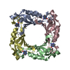
| |||||||||||||||
| Unit cell |
| |||||||||||||||
| Components on special symmetry positions |
|
-Components
| #1: Protein | Mass: 16149.166 Da / Num. of mol.: 4 Source method: isolated from a genetically manipulated source Source: (gene. exp.) Yersinia pestis (bacteria) / Gene: y3531 / Plasmid: pMCSG7 / Production host: References: UniProt: Q8CZR7, UniProt: A0A5P8YJX5*PLUS, dihydroneopterin aldolase #2: Chemical | ChemComp-PE0 /   Mass: 163.137 Da / Num. of mol.: 4 / Source method: obtained synthetically / Formula: C6H5N5O Mass: 163.137 Da / Num. of mol.: 4 / Source method: obtained synthetically / Formula: C6H5N5O#3: Water | ChemComp-HOH / |  Mass: 18.015 Da / Num. of mol.: 437 / Source method: isolated from a natural source / Formula: H2O Mass: 18.015 Da / Num. of mol.: 437 / Source method: isolated from a natural source / Formula: H2O |
|---|
-Experimental details
-Experiment
| Experiment | Method: X-RAY DIFFRACTION / Number of used crystals: 1 |
|---|
- Sample preparation
| Crystal | Density Matthews: 2.17 Å3/Da / Density % sol: 43.22 % |
|---|---|
| Crystal grow | Temperature: 273 K / Method: vapor diffusion, sitting drop Details: 0.1 M BisTris pH 6.5 0.2 M CaCl2 0.05 M NaCl 47% MPD |
-Data collection
| Diffraction | Mean temperature: 100 K / Serial crystal experiment: N |
|---|---|
| Diffraction source | Source: ROTATING ANODE / Type: RIGAKU MICROMAX-007 HF / Wavelength: 1.54 Å |
| Detector | Type: DECTRIS PILATUS 200K / Detector: PIXEL / Date: Jul 1, 2015 |
| Radiation | Protocol: SINGLE WAVELENGTH / Monochromatic (M) / Laue (L): M / Scattering type: x-ray |
| Radiation wavelength | Wavelength: 1.54 Å / Relative weight: 1 |
| Reflection | Resolution: 1.886→41.557 Å / Num. obs: 45556 / % possible obs: 99 % / Redundancy: 4.2 % / Biso Wilson estimate: 19.45 Å2 / Rrim(I) all: 0.11 / Net I/σ(I): 15.5 |
| Reflection shell | Resolution: 1.886→1.954 Å / Redundancy: 2.5 % / Mean I/σ(I) obs: 1.8 / Num. unique obs: 4210 / Rrim(I) all: 0.584 / % possible all: 91.8 |
- Processing
| Software |
| |||||||||||||||||||||||||||||||||||||||||||||||||||||||||||||||||||||||||||||||||||||||||||||||||||||||||
|---|---|---|---|---|---|---|---|---|---|---|---|---|---|---|---|---|---|---|---|---|---|---|---|---|---|---|---|---|---|---|---|---|---|---|---|---|---|---|---|---|---|---|---|---|---|---|---|---|---|---|---|---|---|---|---|---|---|---|---|---|---|---|---|---|---|---|---|---|---|---|---|---|---|---|---|---|---|---|---|---|---|---|---|---|---|---|---|---|---|---|---|---|---|---|---|---|---|---|---|---|---|---|---|---|---|---|
| Refinement | Method to determine structure: MOLECULAR REPLACEMENT Starting model: 3r2e Resolution: 1.886→41.557 Å / SU ML: 0.17 / Cross valid method: THROUGHOUT / σ(F): 1.34 / Phase error: 24.27
| |||||||||||||||||||||||||||||||||||||||||||||||||||||||||||||||||||||||||||||||||||||||||||||||||||||||||
| Solvent computation | Shrinkage radii: 0.9 Å / VDW probe radii: 1.11 Å | |||||||||||||||||||||||||||||||||||||||||||||||||||||||||||||||||||||||||||||||||||||||||||||||||||||||||
| Displacement parameters | Biso max: 77.29 Å2 / Biso mean: 22.2082 Å2 / Biso min: 8.47 Å2 | |||||||||||||||||||||||||||||||||||||||||||||||||||||||||||||||||||||||||||||||||||||||||||||||||||||||||
| Refinement step | Cycle: final / Resolution: 1.886→41.557 Å
| |||||||||||||||||||||||||||||||||||||||||||||||||||||||||||||||||||||||||||||||||||||||||||||||||||||||||
| LS refinement shell | Refine-ID: X-RAY DIFFRACTION / Rfactor Rfree error: 0 / Total num. of bins used: 14
|
