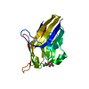
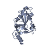
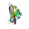
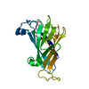
- PDB-6oil: Crystal structure of human VISTA extracellular domain -
+
Open data
ID or keywords:
Loading...
-
Basic information
Entry
Database: PDB / ID: 6oil
Title
Crystal structure of human VISTA extracellular domain
Components
V-type immunoglobulin domain-containing suppressor of T-cell activation
Keywords
SIGNALING PROTEIN / T cell activation / transmembrane / suppressor
Function / homology
Function and homology information
positive regulation of collagen catabolic process / negative regulation of CD8-positive, alpha-beta T cell proliferation / positive regulation of endopeptidase activity / negative regulation of alpha-beta T cell activation / negative regulation of CD4-positive, alpha-beta T cell proliferation / negative regulation of interleukin-17 production / positive regulation of regulatory T cell differentiation / zymogen activation / negative regulation of interleukin-10 production / negative regulation of type II interferon production ...positive regulation of collagen catabolic process / negative regulation of CD8-positive, alpha-beta T cell proliferation / positive regulation of endopeptidase activity / negative regulation of alpha-beta T cell activation / negative regulation of CD4-positive, alpha-beta T cell proliferation / negative regulation of interleukin-17 production / positive regulation of regulatory T cell differentiation / zymogen activation / negative regulation of interleukin-10 production / negative regulation of type II interferon production / negative regulation of tumor necrosis factor production / regulation of immune response / endopeptidase activator activity / positive regulation of cell migration / positive regulation of gene expression / enzyme binding / identical protein binding / plasma membrane Similarity search - Function
Mass: 18.015 Da / Num. of mol.: 41 / Source method: isolated from a natural source / Formula: H2O
Has protein modification
Y
-
Experimental details
-
Experiment
Experiment
Method: X-RAY DIFFRACTION / Number of used crystals: 1
-
Sample preparation
Crystal
Density Matthews: 1.85 Å3/Da / Density % sol: 33.48 %
Crystal grow
Temperature: 293 K / Method: vapor diffusion, sitting drop Details: 18% PEG3350, 0.075M NaBr, 0.05M HAT buffer(combination of HEPES (7.5), ADA (6.5), and TrisHCl (8.0)) PH range: 6.5-8.0
-
Data collection
Diffraction
Mean temperature: 100 K / Serial crystal experiment: N
In the structure databanks used in Yorodumi, some data are registered as the other names, "COVID-19 virus" and "2019-nCoV". Here are the details of the virus and the list of structure data.
Jan 31, 2019. EMDB accession codes are about to change! (news from PDBe EMDB page)
EMDB accession codes are about to change! (news from PDBe EMDB page)
The allocation of 4 digits for EMDB accession codes will soon come to an end. Whilst these codes will remain in use, new EMDB accession codes will include an additional digit and will expand incrementally as the available range of codes is exhausted. The current 4-digit format prefixed with “EMD-” (i.e. EMD-XXXX) will advance to a 5-digit format (i.e. EMD-XXXXX), and so on. It is currently estimated that the 4-digit codes will be depleted around Spring 2019, at which point the 5-digit format will come into force.
The EM Navigator/Yorodumi systems omit the EMD- prefix.
Related info.:Q: What is EMD? / ID/Accession-code notation in Yorodumi/EM Navigator
Yorodumi is a browser for structure data from EMDB, PDB, SASBDB, etc.
This page is also the successor to EM Navigator detail page, and also detail information page/front-end page for Omokage search.
The word "yorodu" (or yorozu) is an old Japanese word meaning "ten thousand". "mi" (miru) is to see.
Related info.:EMDB / PDB / SASBDB / Comparison of 3 databanks / Yorodumi Search / Aug 31, 2016. New EM Navigator & Yorodumi / Yorodumi Papers / Jmol/JSmol / Function and homology information / Changes in new EM Navigator and Yorodumi
 Movie
Movie Controller


 Open data
 Basic information
 Components
 Keywords
 Function and homology information
 Homo sapiens (human)
 X-RAY DIFFRACTION /
  Authors
 United States, 2items
  Citation
 Structure visualization
 Downloads & links
 Other downloads
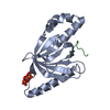


 PDBj
PDBj Assembly
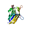

 Type: D-saccharide, beta linking / Mass: 221.208 Da / Num. of mol.: 2
 Mass: 18.015 Da / Num. of mol.: 41 / Source method: isolated from a natural source / Formula: H2O
 Sample preparation
 Processing