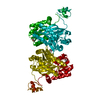
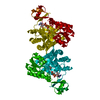
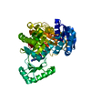
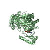
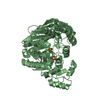
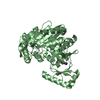
Entry Database : PDB / ID : 6oi8Title Crystal Structure of Haemophilus Influenzae Biotin Carboxylase Complexed with 7-((1R,5S,6s)-6-amino-3-azabicyclo[3.1.0]hexan-3-yl)-6-(2-chloro-6-(pyridin-3-yl)phenyl)pyrido[2,3-d]pyrimidin-2-amine Biotin carboxylase Keywords / / / Function / homology Function Domain/homology Component
/ / / / / / / / / / / / / / / / / / / / / / / / / / / / / / / / / / / / / / / Biological species Haemophilus influenzae (bacteria)Method / / / Resolution : 2.5 Å Authors Andrews, L.D. / Kane, T.R. / Dozzo, P. / Haglund, C.M. / Hilderbrandt, D.J. / Linsell, M.S. / Machajewski, T. / McEnroe, G. / Serio, A.W. / Wlasichuk, K.B. ...Andrews, L.D. / Kane, T.R. / Dozzo, P. / Haglund, C.M. / Hilderbrandt, D.J. / Linsell, M.S. / Machajewski, T. / McEnroe, G. / Serio, A.W. / Wlasichuk, K.B. / Neau, D.B. / Pakhomova, S. / Waldrop, G.L. / Sharp, M. / Pogliano, J. / Cirz, R. / Cohen, F. Funding support Organization Grant number Country National Institutes of Health/National Institute Of Allergy and Infectious Diseases (NIH/NIAID) R21AI113572
Journal : J.Med.Chem. / Year : 2019Title : Optimization and Mechanistic Characterization of Pyridopyrimidine Inhibitors of Bacterial Biotin Carboxylase.Authors: Andrews, L.D. / Kane, T.R. / Dozzo, P. / Haglund, C.M. / Hilderbrandt, D.J. / Linsell, M.S. / Machajewski, T. / McEnroe, G. / Serio, A.W. / Wlasichuk, K.B. / Neau, D.B. / Pakhomova, S. / ... Authors : Andrews, L.D. / Kane, T.R. / Dozzo, P. / Haglund, C.M. / Hilderbrandt, D.J. / Linsell, M.S. / Machajewski, T. / McEnroe, G. / Serio, A.W. / Wlasichuk, K.B. / Neau, D.B. / Pakhomova, S. / Waldrop, G.L. / Sharp, M. / Pogliano, J. / Cirz, R.T. / Cohen, F. History Deposition Apr 9, 2019 Deposition site / Processing site Revision 1.0 Jul 31, 2019 Provider / Type Revision 1.1 Sep 4, 2019 Group / Database references / Category / citation_authorItem _citation.journal_volume / _citation.page_first ... _citation.journal_volume / _citation.page_first / _citation.page_last / _citation_author.name Revision 1.2 Dec 18, 2019 Group / Category / Item Revision 1.3 Oct 11, 2023 Group / Database references / Refinement descriptionCategory chem_comp_atom / chem_comp_bond ... chem_comp_atom / chem_comp_bond / database_2 / pdbx_initial_refinement_model Item / _database_2.pdbx_database_accession
Show all Show less
 Movie
Movie Controller

 Yorodumi
 Open data
 Basic information
 Components
 Keywords
 Function and homology information
 Haemophilus influenzae (bacteria)
 X-RAY DIFFRACTION /
  Authors
 United States, 1items
  Citation
 Structure visualization
 Downloads & links
 Other downloads
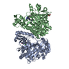


 PDBj
PDBj


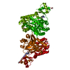
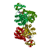
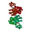
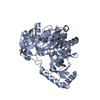
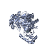
 Assembly

 Mass: 62.068 Da / Num. of mol.: 1 / Source method: obtained synthetically / Formula: C2H6O2
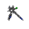
 Mass: 429.905 Da / Num. of mol.: 1 / Source method: obtained synthetically / Formula: C23H20ClN7
 Mass: 18.015 Da / Num. of mol.: 30 / Source method: isolated from a natural source / Formula: H2O
 Sample preparation
 Processing