 Movie
Movie Controller


+ Open data

- Basic information
| Entry | Database: PDB / ID: 6oha | |||||||||
|---|---|---|---|---|---|---|---|---|---|---|
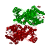
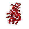
| Title | Yeast Guanine Deaminase | |||||||||
 Components Components | Probable guanine deaminase | |||||||||
 Keywords Keywords | HYDROLASE / amidohydrolase guanine deaminase purine metabolism | |||||||||
| Function / homology |  Function and homology information Function and homology informationPurine catabolism / guanine deaminase / guanine deaminase activity / guanine catabolic process / guanine metabolic process / zinc ion binding / cytoplasm / cytosol Similarity search - Function | |||||||||
| Biological species |  | |||||||||
| Method |  X-RAY DIFFRACTION / SYNCHROTRON / MOLECULAR REPLACEMENT / Resolution: 2.21 Å X-RAY DIFFRACTION / SYNCHROTRON / MOLECULAR REPLACEMENT / Resolution: 2.21 Å | |||||||||
 Authors Authors | Shek, R.S. / French, J.B. | |||||||||
| Funding support |  United States, 2items United States, 2items
| |||||||||
 Citation Citation | Journal: Biochemistry / Year: 2019 Title: Structural Determinants for Substrate Selectivity in Guanine Deaminase Enzymes of the Amidohydrolase Superfamily. Authors: Shek, R. / Hilaire, T. / Sim, J. / French, J.B. | |||||||||
| History |
|
- Structure visualization
| Structure viewer | Molecule: MolmilJmol/JSmol |
|---|
- Downloads & links
-Download
| PDBx/mmCIF format | 6oha.cif.gz | 227 KB | Display | PDBx/mmCIF format |
|---|---|---|---|---|
| PDB format | pdb6oha.ent.gz | 146.4 KB | Display | PDB format |
| PDBx/mmJSON format | 6oha.json.gz | Tree view | PDBx/mmJSON format | |
| Others |  Other downloads Other downloads |
-Validation report
| Arichive directory | https://data.pdbj.org/pub/pdb/validation_reports/oh/6ohaftp://data.pdbj.org/pub/pdb/validation_reports/oh/6oha | HTTPS FTP |
|---|
-Related structure data
| Related structure data | 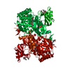 6oh9C  6ohbC  6ohcC  2oodS C: citing same article ( S: Starting model for refinement |
|---|---|
| Similar structure data |


-Links
 PDBj
PDBj


- Assembly
| Deposited unit | 
| ||||||||||||
|---|---|---|---|---|---|---|---|---|---|---|---|---|---|
| 1 | 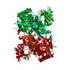
| ||||||||||||
| Unit cell |
| ||||||||||||
| Components on special symmetry positions |
|
-Components
-Protein , 1 types, 1 molecules A
| #1: Protein | Mass: 55269.410 Da / Num. of mol.: 1 Source method: isolated from a genetically manipulated source Source: (gene. exp.) Strain: ATCC 204508 / S288c / Gene: GUD1, YDL238C / Production host:  |
|---|
-Non-polymers , 5 types, 283 molecules 


| #2: Chemical | ChemComp-XAN /  Mass: 152.111 Da / Num. of mol.: 1 / Source method: obtained synthetically / Formula: C5H4N4O2 Mass: 152.111 Da / Num. of mol.: 1 / Source method: obtained synthetically / Formula: C5H4N4O2 | ||||
|---|---|---|---|---|---|
| #3: Chemical | ChemComp-ZN /  Mass: 65.409 Da / Num. of mol.: 1 / Source method: obtained synthetically / Formula: Zn Mass: 65.409 Da / Num. of mol.: 1 / Source method: obtained synthetically / Formula: Zn | ||||
| #4: Chemical |  Mass: 238.278 Da / Num. of mol.: 3 / Source method: obtained synthetically / Formula: C10H22O6 / Comment: precipitant*YM Mass: 238.278 Da / Num. of mol.: 3 / Source method: obtained synthetically / Formula: C10H22O6 / Comment: precipitant*YM#5: Chemical |  Mass: 96.063 Da / Num. of mol.: 3 / Source method: obtained synthetically / Formula: SO4 Mass: 96.063 Da / Num. of mol.: 3 / Source method: obtained synthetically / Formula: SO4#6: Water | ChemComp-HOH / | Mass: 18.015 Da / Num. of mol.: 275 / Source method: isolated from a natural source / Formula: H2O |
-Experimental details
-Experiment
| Experiment | Method: X-RAY DIFFRACTION / Number of used crystals: 1 |
|---|
- Sample preparation
| Crystal | Density Matthews: 3.21 Å3/Da / Density % sol: 61.72 % |
|---|---|
| Crystal grow | Temperature: 291 K / Method: vapor diffusion, hanging drop Details: 17% Peg 3350 0.2 M ammonium sulfate 100 mM Hepes, pH 7.5 |
-Data collection
| Diffraction | Mean temperature: 100 K / Serial crystal experiment: N |
|---|---|
| Diffraction source | Source: SYNCHROTRON / Site: NSLS-II / Beamline: 17-ID-1 / Wavelength: 0.9201 Å |
| Detector | Type: DECTRIS EIGER X 9M / Detector: PIXEL / Date: Aug 15, 2018 |
| Radiation | Protocol: SINGLE WAVELENGTH / Monochromatic (M) / Laue (L): M / Scattering type: x-ray |
| Radiation wavelength | Wavelength: 0.9201 Å / Relative weight: 1 |
| Reflection | Resolution: 2.21→29.07 Å / Num. obs: 33428 / % possible obs: 99.1 % / Redundancy: 8.8 % / Biso Wilson estimate: 32.73 Å2 / CC1/2: 0.996 / Rmerge(I) obs: 0.146 / Net I/σ(I): 10 |
| Reflection shell | Resolution: 2.21→2.29 Å / Rmerge(I) obs: 0.973 / Num. unique obs: 3075 / CC1/2: 0.696 |
- Processing
| Software |
| |||||||||||||||||||||||||||||||||||||||||||||||||||||||||||||||||||||||||||||||||||||||||||
|---|---|---|---|---|---|---|---|---|---|---|---|---|---|---|---|---|---|---|---|---|---|---|---|---|---|---|---|---|---|---|---|---|---|---|---|---|---|---|---|---|---|---|---|---|---|---|---|---|---|---|---|---|---|---|---|---|---|---|---|---|---|---|---|---|---|---|---|---|---|---|---|---|---|---|---|---|---|---|---|---|---|---|---|---|---|---|---|---|---|---|---|---|
| Refinement | Method to determine structure: MOLECULAR REPLACEMENT Starting model: 2OOD Resolution: 2.21→29.07 Å / SU ML: 0.2169 / Cross valid method: FREE R-VALUE / σ(F): 1.35 / Phase error: 20.2395
| |||||||||||||||||||||||||||||||||||||||||||||||||||||||||||||||||||||||||||||||||||||||||||
| Solvent computation | Shrinkage radii: 0.9 Å / VDW probe radii: 1.11 Å | |||||||||||||||||||||||||||||||||||||||||||||||||||||||||||||||||||||||||||||||||||||||||||
| Displacement parameters | Biso mean: 34.42 Å2 | |||||||||||||||||||||||||||||||||||||||||||||||||||||||||||||||||||||||||||||||||||||||||||
| Refinement step | Cycle: LAST / Resolution: 2.21→29.07 Å
| |||||||||||||||||||||||||||||||||||||||||||||||||||||||||||||||||||||||||||||||||||||||||||
| Refine LS restraints |
| |||||||||||||||||||||||||||||||||||||||||||||||||||||||||||||||||||||||||||||||||||||||||||
| LS refinement shell |
|
