 Movie
Movie Controller

[English] 日本語
 Yorodumi
- PDB-6of8: Structure of Thr354Asn, Glu355Gln, Thr412Asn, Ile414Met, Ile464Hi... -
+ Open data

- Basic information
| Entry | Database: PDB / ID: 6of8 | |||||||||
|---|---|---|---|---|---|---|---|---|---|---|
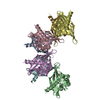
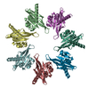
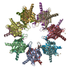
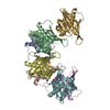
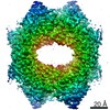
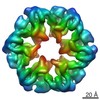
| Title | Structure of Thr354Asn, Glu355Gln, Thr412Asn, Ile414Met, Ile464His, and Phe467Met mutant human CamKII-alpha hub domain | |||||||||
 Components Components | Calcium/calmodulin-dependent protein kinase type II subunit alpha | |||||||||
 Keywords Keywords | TRANSFERASE / CaMKII hub domain / SIGNALING PROTEIN | |||||||||
| Function / homology |  Function and homology information Function and homology informationpeptidyl-threonine autophosphorylation / calcium- and calmodulin-dependent protein kinase complex / regulation of endocannabinoid signaling pathway / Ca2+/calmodulin-dependent protein kinase / dendritic spine development / negative regulation of hydrolase activity / regulation of neurotransmitter secretion / Trafficking of AMPA receptors / positive regulation of calcium ion transport / regulation of neuron migration ...peptidyl-threonine autophosphorylation / calcium- and calmodulin-dependent protein kinase complex / regulation of endocannabinoid signaling pathway / Ca2+/calmodulin-dependent protein kinase / dendritic spine development / negative regulation of hydrolase activity / regulation of neurotransmitter secretion / Trafficking of AMPA receptors / positive regulation of calcium ion transport / regulation of neuron migration / calcium/calmodulin-dependent protein kinase activity / regulation of mitochondrial membrane permeability involved in apoptotic process / Assembly and cell surface presentation of NMDA receptors / CaMK IV-mediated phosphorylation of CREB / Phase 0 - rapid depolarisation / Negative regulation of NMDA receptor-mediated neuronal transmission / Unblocking of NMDA receptors, glutamate binding and activation / negative regulation of ferroptosis / Ion transport by P-type ATPases / Long-term potentiation / HSF1-dependent transactivation / Regulation of MECP2 expression and activity / regulation of neuronal synaptic plasticity / glutamate receptor binding / regulation of protein localization to plasma membrane / cellular response to interferon-beta / Ion homeostasis / positive regulation of cardiac muscle cell apoptotic process / Ras activation upon Ca2+ influx through NMDA receptor / response to ischemia / : / angiotensin-activated signaling pathway / positive regulation of receptor signaling pathway via JAK-STAT / G1/S transition of mitotic cell cycle / RAF activation / cellular response to type II interferon / Interferon gamma signaling / kinase activity / Signaling by RAF1 mutants / Signaling by moderate kinase activity BRAF mutants / Paradoxical activation of RAF signaling by kinase inactive BRAF / Signaling downstream of RAS mutants / calcium ion transport / endocytic vesicle membrane / Signaling by BRAF and RAF1 fusions / long-term synaptic potentiation / RAF/MAP kinase cascade / Ca2+ pathway / dendritic spine / protein phosphorylation / calmodulin binding / neuron projection / postsynaptic density / protein serine kinase activity / protein serine/threonine kinase activity / protein homodimerization activity / mitochondrion / nucleoplasm / ATP binding / metal ion binding / identical protein binding / nucleus / cytoplasm / cytosol Similarity search - Function | |||||||||
| Biological species |  Homo sapiens (human) Homo sapiens (human) | |||||||||
| Method |  X-RAY DIFFRACTION / SYNCHROTRON / MOLECULAR REPLACEMENT / Resolution: 2.1 Å X-RAY DIFFRACTION / SYNCHROTRON / MOLECULAR REPLACEMENT / Resolution: 2.1 Å | |||||||||
 Authors Authors | McSpadden, E.D. / Chi, C.C. / Gee, C.L. / Kuriyan, J. | |||||||||
| Funding support |  United States, 2items United States, 2items
| |||||||||
 Citation Citation | Journal: Protein Sci. / Year: 2019 Title: Variation in assembly stoichiometry in non-metazoan homologs of the hub domain of Ca2+/calmodulin-dependent protein kinase II. Authors: McSpadden, E.D. / Xia, Z. / Chi, C.C. / Susa, A.C. / Shah, N.H. / Gee, C.L. / Williams, E.R. / Kuriyan, J. | |||||||||
| History |
|
- Structure visualization
| Structure viewer | Molecule: MolmilJmol/JSmol |
|---|
- Downloads & links
-Download
| PDBx/mmCIF format | 6of8.cif.gz | 197 KB | Display | PDBx/mmCIF format |
|---|---|---|---|---|
| PDB format | pdb6of8.ent.gz | 157.7 KB | Display | PDB format |
| PDBx/mmJSON format | 6of8.json.gz | Tree view | PDBx/mmJSON format | |
| Others |  Other downloads Other downloads |
-Validation report
| Arichive directory | https://data.pdbj.org/pub/pdb/validation_reports/of/6of8ftp://data.pdbj.org/pub/pdb/validation_reports/of/6of8 | HTTPS FTP |
|---|
-Related structure data
| Related structure data |  6of9C 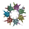 1hkxS S: Starting model for refinement C: citing same article ( |
|---|---|
| Similar structure data |
-Links
 PDBj
PDBj


- Assembly
| Deposited unit | 
| ||||||||
|---|---|---|---|---|---|---|---|---|---|
| 1 | 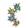
| ||||||||
| Unit cell |
|
-Components
| #1: Protein | Mass: 15551.578 Da / Num. of mol.: 7 / Mutation: T354N, E355Q, T412N, I414M, I464H, F467M Source method: isolated from a genetically manipulated source Source: (gene. exp.) Homo sapiens (human) / Gene: CAMK2A, CAMKA, KIAA0968 / Production host:  References: UniProt: Q9UQM7, Ca2+/calmodulin-dependent protein kinase #2: Chemical | ChemComp-K /   Mass: 39.098 Da / Num. of mol.: 5 / Source method: isolated from a natural source / Formula: K Mass: 39.098 Da / Num. of mol.: 5 / Source method: isolated from a natural source / Formula: K#3: Chemical |   Mass: 92.094 Da / Num. of mol.: 3 / Source method: obtained synthetically / Formula: C3H8O3 Mass: 92.094 Da / Num. of mol.: 3 / Source method: obtained synthetically / Formula: C3H8O3#4: Water | ChemComp-HOH / |  Mass: 18.015 Da / Num. of mol.: 105 / Source method: isolated from a natural source / Formula: H2O Mass: 18.015 Da / Num. of mol.: 105 / Source method: isolated from a natural source / Formula: H2O |
|---|
-Experimental details
-Experiment
| Experiment | Method: X-RAY DIFFRACTION / Number of used crystals: 1 |
|---|
- Sample preparation
| Crystal | Density Matthews: 2.43 Å3/Da / Density % sol: 49.32 % |
|---|---|
| Crystal grow | Temperature: 293 K / Method: vapor diffusion, sitting drop Details: 17 mg/mL mutant hub in 25 mM Tris, 150 mM KCl, 10% (v/v) glycerol, 2 mM DTT, 1 mM TCEP, pH 8.0 was mixed 1:1 with 235 mM K3Citrate, 15% (w/v) PEG 3350, pH 8.0 |
-Data collection
| Diffraction | Mean temperature: 100 K / Serial crystal experiment: N | ||||||||||||||||||||||||
|---|---|---|---|---|---|---|---|---|---|---|---|---|---|---|---|---|---|---|---|---|---|---|---|---|---|
| Diffraction source | Source: SYNCHROTRON / Site: ALS / Beamline: 8.3.1 / Wavelength: 1.11583 Å | ||||||||||||||||||||||||
| Detector | Type: DECTRIS PILATUS3 6M / Detector: PIXEL / Date: Nov 11, 2017 | ||||||||||||||||||||||||
| Radiation | Monochromator: S111 / Protocol: SINGLE WAVELENGTH / Monochromatic (M) / Laue (L): M / Scattering type: x-ray | ||||||||||||||||||||||||
| Radiation wavelength | Wavelength: 1.11583 Å / Relative weight: 1 | ||||||||||||||||||||||||
| Reflection | Resolution: 2.1→47.76 Å / Num. obs: 60334 / % possible obs: 99.5 % / Redundancy: 3.1 % / CC1/2: 0.999 / Rmerge(I) obs: 0.039 / Rpim(I) all: 0.026 / Rrim(I) all: 0.047 / Net I/σ(I): 13 | ||||||||||||||||||||||||
| Reflection shell | Diffraction-ID: 1 / Redundancy: 3.1 %
|
- Processing
| Software |
| |||||||||||||||||||||||||||||||||||||||||||||||||||||||||||||||||||||||||||||||||||||||||||||||||||||||||||||||||||||||||||||||||||||||||||||||||||||||||||||||||
|---|---|---|---|---|---|---|---|---|---|---|---|---|---|---|---|---|---|---|---|---|---|---|---|---|---|---|---|---|---|---|---|---|---|---|---|---|---|---|---|---|---|---|---|---|---|---|---|---|---|---|---|---|---|---|---|---|---|---|---|---|---|---|---|---|---|---|---|---|---|---|---|---|---|---|---|---|---|---|---|---|---|---|---|---|---|---|---|---|---|---|---|---|---|---|---|---|---|---|---|---|---|---|---|---|---|---|---|---|---|---|---|---|---|---|---|---|---|---|---|---|---|---|---|---|---|---|---|---|---|---|---|---|---|---|---|---|---|---|---|---|---|---|---|---|---|---|---|---|---|---|---|---|---|---|---|---|---|---|---|---|---|---|
| Refinement | Method to determine structure: MOLECULAR REPLACEMENT Starting model: 1HKX Resolution: 2.1→47.505 Å / SU ML: 0.32 / Cross valid method: FREE R-VALUE / σ(F): 1.34 / Phase error: 30.96 / Stereochemistry target values: ML
| |||||||||||||||||||||||||||||||||||||||||||||||||||||||||||||||||||||||||||||||||||||||||||||||||||||||||||||||||||||||||||||||||||||||||||||||||||||||||||||||||
| Solvent computation | Shrinkage radii: 0.9 Å / VDW probe radii: 1.11 Å / Solvent model: FLAT BULK SOLVENT MODEL | |||||||||||||||||||||||||||||||||||||||||||||||||||||||||||||||||||||||||||||||||||||||||||||||||||||||||||||||||||||||||||||||||||||||||||||||||||||||||||||||||
| Refinement step | Cycle: LAST / Resolution: 2.1→47.505 Å
| |||||||||||||||||||||||||||||||||||||||||||||||||||||||||||||||||||||||||||||||||||||||||||||||||||||||||||||||||||||||||||||||||||||||||||||||||||||||||||||||||
| Refine LS restraints |
| |||||||||||||||||||||||||||||||||||||||||||||||||||||||||||||||||||||||||||||||||||||||||||||||||||||||||||||||||||||||||||||||||||||||||||||||||||||||||||||||||
| LS refinement shell |
|
