 Movie
Movie Controller


+ Open data

- Basic information
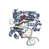
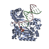
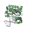
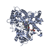
| Entry | Database: PDB / ID: 6oaj | ||||||
|---|---|---|---|---|---|---|---|
| Title | HUaE34K 19bp SYM DNA | ||||||
 Components Components |
| ||||||
 Keywords Keywords | dna binding protein/dna / Nucleoid Associated Protein / DNA supercoiling / Histone like proteins / DNA BINDING PROTEIN / dna binding protein-dna complex | ||||||
| Function / homology |  Function and homology information Function and homology informationHU-DNA complex / bacterial nucleoid DNA packaging / DnaA-HU complex / chromosome condensation / DNA replication initiation / structural constituent of chromatin / DNA repair / DNA damage response / DNA-templated transcription / DNA binding ...HU-DNA complex / bacterial nucleoid DNA packaging / DnaA-HU complex / chromosome condensation / DNA replication initiation / structural constituent of chromatin / DNA repair / DNA damage response / DNA-templated transcription / DNA binding / membrane / identical protein binding / cytosol Similarity search - Function | ||||||
| Biological species |  | ||||||
| Method |  X-RAY DIFFRACTION / SYNCHROTRON / MOLECULAR REPLACEMENT / Resolution: 4.092 Å X-RAY DIFFRACTION / SYNCHROTRON / MOLECULAR REPLACEMENT / Resolution: 4.092 Å | ||||||
 Authors Authors | Remesh, S.G. / Hammel, M. | ||||||
| Funding support |  United States, 1items United States, 1items
| ||||||
 Citation Citation | Journal: Nat Commun / Year: 2020 Title: Nucleoid remodeling during environmental adaptation is regulated by HU-dependent DNA bundling. Authors: Remesh, S.G. / Verma, S.C. / Chen, J.H. / Ekman, A.A. / Larabell, C.A. / Adhya, S. / Hammel, M. | ||||||
| History |
|
- Structure visualization
| Structure viewer | Molecule: MolmilJmol/JSmol |
|---|
- Downloads & links
-Download
| PDBx/mmCIF format | 6oaj.cif.gz | 78.7 KB | Display | PDBx/mmCIF format |
|---|---|---|---|---|
| PDB format | pdb6oaj.ent.gz | 54.5 KB | Display | PDB format |
| PDBx/mmJSON format | 6oaj.json.gz | Tree view | PDBx/mmJSON format | |
| Others |  Other downloads Other downloads |
-Validation report
| Arichive directory | https://data.pdbj.org/pub/pdb/validation_reports/oa/6oajftp://data.pdbj.org/pub/pdb/validation_reports/oa/6oaj | HTTPS FTP |
|---|
-Related structure data
| Related structure data | 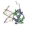 6o6kC 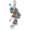 6o8qC 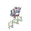 4yexS S: Starting model for refinement C: citing same article ( |
|---|---|
| Similar structure data |


-Links
 PDBj
PDBj


- Assembly
| Deposited unit | 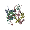
| ||||||||
|---|---|---|---|---|---|---|---|---|---|
| 1 |
| ||||||||
| Unit cell |
|
-Components
| #1: Protein | Mass: 9550.046 Da / Num. of mol.: 4 / Mutation: E34K Source method: isolated from a genetically manipulated source Source: (gene. exp.) Strain: K12 / Gene: hupA, b4000, JW3964 Production host: Strain (production host): BL21-Gold(DE3)pLysS AG / References: UniProt: P0ACF0 #2: DNA chain | | Mass: 5821.759 Da / Num. of mol.: 1 / Source method: obtained synthetically / Source: (synth.) #3: DNA chain | | Mass: 6119.956 Da / Num. of mol.: 1 / Source method: obtained synthetically / Source: (synth.) |
|---|
-Experimental details
-Experiment
| Experiment | Method: X-RAY DIFFRACTION / Number of used crystals: 1 |
|---|
- Sample preparation
| Crystal | Density Matthews: 2.55 Å3/Da / Density % sol: 51.77 % |
|---|---|
| Crystal grow | Temperature: 293 K / Method: vapor diffusion, hanging drop / pH: 5 / Details: 0.1M Na-malonate, pH 5.0, 12% PEG 3350 |
-Data collection
| Diffraction | Mean temperature: 100 K / Serial crystal experiment: N |
|---|---|
| Diffraction source | Source: SYNCHROTRON / Site: ALS / Beamline: 8.3.1 / Wavelength: 1.11582 Å |
| Detector | Type: DECTRIS PILATUS3 S 6M / Detector: PIXEL / Date: Sep 14, 2016 |
| Radiation | Protocol: SINGLE WAVELENGTH / Monochromatic (M) / Laue (L): M / Scattering type: x-ray |
| Radiation wavelength | Wavelength: 1.11582 Å / Relative weight: 1 |
| Reflection | Resolution: 4.092→40.492 Å / Num. obs: 4331 / % possible obs: 99.29 % / Redundancy: 6.2 % / CC1/2: 0.996 / Rmerge(I) obs: 0.119 / Rpim(I) all: 0.05499 / Rrim(I) all: 0.1316 / Net I/σ(I): 7.28 |
| Reflection shell | Resolution: 4.092→4.238 Å / Redundancy: 6 % / Rmerge(I) obs: 1.942 / Mean I/σ(I) obs: 1.01 / Num. unique obs: 406 / CC1/2: 0.479 / Rrim(I) all: 2.129 / % possible all: 98.54 |
- Processing
| Software |
| |||||||||||||||||||||||||||||||||||||||||||||||||
|---|---|---|---|---|---|---|---|---|---|---|---|---|---|---|---|---|---|---|---|---|---|---|---|---|---|---|---|---|---|---|---|---|---|---|---|---|---|---|---|---|---|---|---|---|---|---|---|---|---|---|
| Refinement | Method to determine structure: MOLECULAR REPLACEMENT Starting model: 4YEX Resolution: 4.092→40.492 Å / SU ML: 1.09 / Cross valid method: FREE R-VALUE / σ(F): 1.34 / Phase error: 48.42 / Stereochemistry target values: ML
| |||||||||||||||||||||||||||||||||||||||||||||||||
| Solvent computation | Shrinkage radii: 0.9 Å / VDW probe radii: 1.11 Å / Solvent model: FLAT BULK SOLVENT MODEL | |||||||||||||||||||||||||||||||||||||||||||||||||
| Refinement step | Cycle: LAST / Resolution: 4.092→40.492 Å
| |||||||||||||||||||||||||||||||||||||||||||||||||
| Refine LS restraints |
| |||||||||||||||||||||||||||||||||||||||||||||||||
| LS refinement shell |
|
