 Movie
Movie Controller


+ Open data

- Basic information
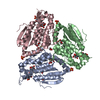
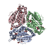
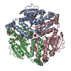
| Entry | Database: PDB / ID: 6ny9 | ||||||
|---|---|---|---|---|---|---|---|
| Title | Alpha/beta hydrolase domain-containing protein 10 from mouse | ||||||
 Components Components | Mycophenolic acid acyl-glucuronide esterase, mitochondrial | ||||||
 Keywords Keywords | HYDROLASE / Alpha/beta hydrolase / Depalmitoylase / Mitochondria | ||||||
| Function / homology |  Function and homology information Function and homology informationGlucuronidation / mycophenolic acid acyl-glucuronide esterase activity / mycophenolic acid acyl-glucuronide esterase / protein depalmitoylation / palmitoyl[protein] hydrolase / palmitoyl-(protein) hydrolase activity / glucuronoside catabolic process / hydrolase activity, hydrolyzing O-glycosyl compounds / mitochondrion / cytosol Similarity search - Function | ||||||
| Biological species |  | ||||||
| Method |  X-RAY DIFFRACTION / SYNCHROTRON / MOLECULAR REPLACEMENT / Resolution: 1.66 Å X-RAY DIFFRACTION / SYNCHROTRON / MOLECULAR REPLACEMENT / Resolution: 1.66 Å | ||||||
 Authors Authors | Cao, Y. / Rice, P.A. / Dickinson, B.C. | ||||||
| Funding support |  United States, 1items United States, 1items
| ||||||
 Citation Citation | Journal: Nat.Chem.Biol. / Year: 2019 Title: ABHD10 is an S-depalmitoylase affecting redox homeostasis through peroxiredoxin-5. Authors: Cao, Y. / Qiu, T. / Kathayat, R.S. / Azizi, S.A. / Thorne, A.K. / Ahn, D. / Fukata, Y. / Fukata, M. / Rice, P.A. / Dickinson, B.C. | ||||||
| History |
|
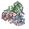
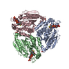
- Structure visualization
| Structure viewer | Molecule: MolmilJmol/JSmol |
|---|
- Downloads & links
-Download
| PDBx/mmCIF format | 6ny9.cif.gz | 194.1 KB | Display | PDBx/mmCIF format |
|---|---|---|---|---|
| PDB format | pdb6ny9.ent.gz | 131.6 KB | Display | PDB format |
| PDBx/mmJSON format | 6ny9.json.gz | Tree view | PDBx/mmJSON format | |
| Others |  Other downloads Other downloads |
-Validation report
| Arichive directory | https://data.pdbj.org/pub/pdb/validation_reports/ny/6ny9ftp://data.pdbj.org/pub/pdb/validation_reports/ny/6ny9 | HTTPS FTP |
|---|
-Related structure data
| Related structure data | 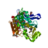 3llcS S: Starting model for refinement |
|---|---|
| Similar structure data |


-Links
 PDBj
PDBj


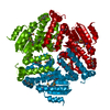
- Assembly
| Deposited unit | 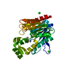
| ||||||||||||
|---|---|---|---|---|---|---|---|---|---|---|---|---|---|
| 1 | 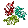
| ||||||||||||
| Unit cell |
| ||||||||||||
| Components on special symmetry positions |
|
-Components
| #1: Protein | Mass: 27821.311 Da / Num. of mol.: 1 Source method: isolated from a genetically manipulated source Source: (gene. exp.)  References: UniProt: Q6PE15, mycophenolic acid acyl-glucuronide esterase | ||
|---|---|---|---|
| #2: Chemical | ChemComp-MPD / (  Mass: 118.174 Da / Num. of mol.: 1 / Source method: obtained synthetically / Formula: C6H14O2 / Comment: precipitant*YM Mass: 118.174 Da / Num. of mol.: 1 / Source method: obtained synthetically / Formula: C6H14O2 / Comment: precipitant*YM | ||
| #3: Chemical | ChemComp-SR /   Mass: 87.620 Da / Num. of mol.: 4 / Source method: obtained synthetically / Formula: Sr Mass: 87.620 Da / Num. of mol.: 4 / Source method: obtained synthetically / Formula: Sr#4: Water | ChemComp-HOH / |  Mass: 18.015 Da / Num. of mol.: 205 / Source method: isolated from a natural source / Formula: H2O Mass: 18.015 Da / Num. of mol.: 205 / Source method: isolated from a natural source / Formula: H2O |
-Experimental details
-Experiment
| Experiment | Method: X-RAY DIFFRACTION / Number of used crystals: 1 |
|---|
- Sample preparation
| Crystal | Density Matthews: 3.83 Å3/Da / Density % sol: 67.91 % |
|---|---|
| Crystal grow | Temperature: 277.15 K / Method: liquid diffusion / pH: 6.4 Details: 0.08 M strontium chloride hexahydrate, 0.04 M sodium cacodylate trihydrate pH 6.4, 25% v/v (+/-)-2-methyl-2,4-pentanediol (MPD), 0.012 M spermine tetrahydrochloride PH range: 6.0-6.4 |
-Data collection
| Diffraction | Mean temperature: 100 K / Serial crystal experiment: N |
|---|---|
| Diffraction source | Source: SYNCHROTRON / Site: APS / Beamline: 24-ID-C / Wavelength: 0.979 Å |
| Detector | Type: DECTRIS PILATUS 6M-F / Detector: PIXEL / Date: Nov 14, 2017 |
| Radiation | Protocol: SINGLE WAVELENGTH / Monochromatic (M) / Laue (L): M / Scattering type: x-ray |
| Radiation wavelength | Wavelength: 0.979 Å / Relative weight: 1 |
| Reflection | Resolution: 1.657→56.46 Å / Num. obs: 52270 / % possible obs: 92.82 % / Redundancy: 12.9 % / Biso Wilson estimate: 16.57 Å2 / CC1/2: 0.998 / Rmerge(I) obs: 0.1082 / Net I/σ(I): 20.82 |
| Reflection shell | Resolution: 1.657→1.717 Å / Redundancy: 12.8 % / Mean I/σ(I) obs: 1.6 / Num. unique obs: 4994 / CC1/2: 0.686 / % possible all: 76.98 |
- Processing
| Software |
| ||||||||||||||||||||||||||||||||||||||||||||||||||||||||||||||||||||||||||||||||||||||||||||||||||
|---|---|---|---|---|---|---|---|---|---|---|---|---|---|---|---|---|---|---|---|---|---|---|---|---|---|---|---|---|---|---|---|---|---|---|---|---|---|---|---|---|---|---|---|---|---|---|---|---|---|---|---|---|---|---|---|---|---|---|---|---|---|---|---|---|---|---|---|---|---|---|---|---|---|---|---|---|---|---|---|---|---|---|---|---|---|---|---|---|---|---|---|---|---|---|---|---|---|---|---|
| Refinement | Method to determine structure: MOLECULAR REPLACEMENT Starting model: 3LLC Resolution: 1.66→56.46 Å / SU ML: 0.1917 / Cross valid method: FREE R-VALUE / σ(F): 0 / Phase error: 22.0141
| ||||||||||||||||||||||||||||||||||||||||||||||||||||||||||||||||||||||||||||||||||||||||||||||||||
| Solvent computation | Shrinkage radii: 0.9 Å / VDW probe radii: 1.11 Å | ||||||||||||||||||||||||||||||||||||||||||||||||||||||||||||||||||||||||||||||||||||||||||||||||||
| Displacement parameters | Biso mean: 24.19 Å2 | ||||||||||||||||||||||||||||||||||||||||||||||||||||||||||||||||||||||||||||||||||||||||||||||||||
| Refinement step | Cycle: LAST / Resolution: 1.66→56.46 Å
| ||||||||||||||||||||||||||||||||||||||||||||||||||||||||||||||||||||||||||||||||||||||||||||||||||
| Refine LS restraints |
| ||||||||||||||||||||||||||||||||||||||||||||||||||||||||||||||||||||||||||||||||||||||||||||||||||
| LS refinement shell |
| ||||||||||||||||||||||||||||||||||||||||||||||||||||||||||||||||||||||||||||||||||||||||||||||||||
| Refinement TLS params. | Method: refined / Origin x: 29.4150756586 Å / Origin y: -34.2369923944 Å / Origin z: 17.0271591659 Å
| ||||||||||||||||||||||||||||||||||||||||||||||||||||||||||||||||||||||||||||||||||||||||||||||||||
| Refinement TLS group | Selection details: all |
