[English] 日本語
Yorodumi
- PDB-6ny0: Crystal structure of trimethoprim-resistant type II dihydrofolate... -

+
Open data


ID or keywords:

Loading...

-
Basic information

Entry
Database: PDB / ID: 6ny0
TitleCrystal structure of trimethoprim-resistant type II dihydrofolate reductase in complex with a bisbenzimidazole inhibitor
ComponentsDihydrofolate reductase type 2
KeywordsOXIDOREDUCTASE / antibiotic resistance / selective inhibitor / R67 DHFR / SH3-like barrel / ANTIBIOTIC
Function / homology
Function and homology information


response to methotrexate / dihydrofolate reductase / dihydrofolate reductase activity / tetrahydrofolate biosynthetic process / one-carbon metabolic process / response to xenobiotic stimulus / response to antibiotic
Similarity search - Function
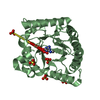
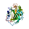
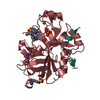
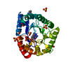
Dihydrofolate reductase, type II / R67 dihydrofolate reductase / SH3 type barrels. - #60 / Mechanosensitive ion channel MscS, beta-domain superfamily / Electron transport accessory-like domain superfamily / SH3 type barrels. / Roll / Mainly Beta
Similarity search - Domain/homology
Chem-LBA / PHOSPHATE ION / Dihydrofolate reductase type 2
Similarity search - Component
Biological speciesEscherichia coli (E. coli)
MethodX-RAY DIFFRACTION / SYNCHROTRON / MOLECULAR REPLACEMENT / Resolution: 1.4 Å
AuthorsYachnin, B.J. / Berghuis, A.M.
Funding support Canada, 5items
OrganizationGrant numberCountry
Natural Sciences and Engineering Research Council (NSERC, Canada)227853 Canada
Natural Sciences and Engineering Research Council (NSERC, Canada)2018-04686 Canada
Other governmentCanada Foundation for Innovation 11510 (www.innovation.ca) Canada
Canadian Institutes of Health Research (CIHR)MOP-13107 Canada
Other privateFondation Marcel et Rolande Gosselin Canada
CitationJournal: Acs Omega / Year: 2019
Title: Structure-Based Design of Dimeric Bisbenzimidazole Inhibitors to an Emergent Trimethoprim-Resistant Type II Dihydrofolate Reductase Guides the Design of Monomeric Analogues.
Authors: Toulouse, J.L. / Yachnin, B.J. / Ruediger, E.H. / Deon, D. / Gagnon, M. / Saint-Jacques, K. / Ebert, M.C.C.J.C. / Forge, D. / Bastien, D. / Colin, D.Y. / Vanden Eynde, J.J. / Marinier, A. / ...Authors: Toulouse, J.L. / Yachnin, B.J. / Ruediger, E.H. / Deon, D. / Gagnon, M. / Saint-Jacques, K. / Ebert, M.C.C.J.C. / Forge, D. / Bastien, D. / Colin, D.Y. / Vanden Eynde, J.J. / Marinier, A. / Berghuis, A.M. / Pelletier, J.N.
History
DepositionFeb 10, 2019Deposition site: RCSB / Processing site: RCSB
Revision 1.0May 29, 2019Provider: repository / Type: Initial release
Revision 1.1Jul 3, 2019Group: Data collection / Database references / Category: citation
Item: _citation.country / _citation.journal_abbrev ..._citation.country / _citation.journal_abbrev / _citation.journal_id_CSD / _citation.journal_id_ISSN / _citation.pdbx_database_id_DOI / _citation.title / _citation.year
Revision 1.2Sep 11, 2019Group: Data collection / Database references / Category: citation / citation_author
Item: _citation.journal_volume / _citation.page_first ..._citation.journal_volume / _citation.page_first / _citation.page_last / _citation.pdbx_database_id_PubMed / _citation.title
Revision 1.3Jan 8, 2020Group: Author supporting evidence / Category: pdbx_audit_support / Item: _pdbx_audit_support.funding_organization
Revision 1.4Oct 11, 2023Group: Data collection / Database references / Refinement description
Category: chem_comp_atom / chem_comp_bond ...chem_comp_atom / chem_comp_bond / database_2 / pdbx_initial_refinement_model
Item: _database_2.pdbx_DOI / _database_2.pdbx_database_accession

-
Structure visualization

Structure viewerMolecule:
MolmilJmol/JSmol

Downloads & links

-
Assembly

Deposited unit
A: Dihydrofolate reductase type 2
hetero molecules


Theoretical massNumber of molelcules
Total (without water)7,6256
Polymers6,6341
Non-polymers9915
Water1,15364
1
A: Dihydrofolate reductase type 2
hetero molecules

A: Dihydrofolate reductase type 2
hetero molecules

A: Dihydrofolate reductase type 2
hetero molecules

A: Dihydrofolate reductase type 2
hetero molecules


Theoretical massNumber of molelcules
Total (without water)30,50124
Polymers26,5384
Non-polymers3,96320
Water724
TypeNameSymmetry operationNumber
identity operation1_555x,y,z1
crystal symmetry operation8_555-y,-x,-z1
crystal symmetry operation10_555-x,-y,z1
crystal symmetry operation15_555y,x,-z1
Buried area7310 Å2
ΔGint-59 kcal/mol
Surface area11650 Å2
MethodPISA
Unit cell
Length a, b, c (Å)67.492, 67.492, 51.791
Angle α, β, γ (deg.)90.000, 90.000, 90.000
Int Tables number98
Space group name H-MI4122

-
Components

#1: Protein Dihydrofolate reductase type 2 / Dihydrofolate reductase type II


Mass: 6634.382 Da / Num. of mol.: 1
Source method: isolated from a genetically manipulated source
Source: (gene. exp.) Escherichia coli (E. coli) / Production host: Escherichia coli BL21 (bacteria) / Strain (production host): BL21 / Variant (production host): pRep4 / References: UniProt: P00383, dihydrofolate reductase
#2: Chemical ChemComp-LBA / 2-(4-{3-[4-(6-carboxy-1H-benzimidazol-2-yl)phenoxy]-2-hydroxypropoxy}phenyl)-1H-benzimidazole-5-carboxylic acid


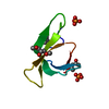
Mass: 564.545 Da / Num. of mol.: 1 / Source method: obtained synthetically / Formula: C31H24N4O7 / Feature type: SUBJECT OF INVESTIGATION
#3: Chemical ChemComp-MRD / (4R)-2-METHYLPENTANE-2,4-DIOL / 2-Methyl-2,4-pentanediol


Mass: 118.174 Da / Num. of mol.: 2 / Source method: obtained synthetically / Formula: C6H14O2 / Comment: precipitant*YM
#4: Chemical ChemComp-PO4 / PHOSPHATE ION / Phosphate


Mass: 94.971 Da / Num. of mol.: 2 / Source method: obtained synthetically / Formula: PO4
#5: Water ChemComp-HOH / water / Water


Mass: 18.015 Da / Num. of mol.: 64 / Source method: isolated from a natural source / Formula: H2O

-
Experimental details

-
Experiment

ExperimentMethod: X-RAY DIFFRACTION / Number of used crystals: 1

-
Sample preparation

CrystalDensity Matthews: 2.22 Å3/Da / Density % sol: 44.66 %
Crystal growTemperature: 277 K / Method: vapor diffusion, hanging drop / pH: 8
Details: The protein was concentrated to 20 mg/mL in 100 mM Tris pH 8.0. Immediately before crystallization, chymotrypsin was added to the sample in a ratio of 1:100 chymotrypsin:protein, and the ...Details: The protein was concentrated to 20 mg/mL in 100 mM Tris pH 8.0. Immediately before crystallization, chymotrypsin was added to the sample in a ratio of 1:100 chymotrypsin:protein, and the protein was diluted to 15 mg/mL using MPD, resulting in a final MPD concentration of 25%. Reservoirs were prepared using 750 uL of 100 mM sodium phosphate pH 7.6 and 60% MPD in a Greiner 24-well hanging-drop crystallization plate. On a siliconized glass cover slip (Hampton Research), 1.5 uL of protein were combined with 2.5 uL of reservoir solution and suspended over the well. The plate was incubated at 277 K and crystals were obtained in a few days.

-
Data collection

DiffractionMean temperature: 93 K / Serial crystal experiment: N
Diffraction sourceSource: SYNCHROTRON / Site: CLSI / Beamline: 08ID-1 / Wavelength: 0.9795 Å
DetectorType: RAYONIX MX300HE / Detector: CCD / Date: Aug 12, 2011
Details: Vertical Focusing Mirror: ultra-low expansion (ULE) titanium siliicate flat mirror with Pt, Uncoated, and Pd strips
RadiationMonochromator: ACCEL/BRUKER double crystal monochromator (DCM), featuring indirectly cryo-cooled first crystal and sagittally focusing second crystal
Protocol: SINGLE WAVELENGTH / Monochromatic (M) / Laue (L): M / Scattering type: x-ray
Radiation wavelengthWavelength: 0.9795 Å / Relative weight: 1
ReflectionResolution: 1.35→50 Å / Num. obs: 13545 / % possible obs: 100 % / Redundancy: 20.1 % / Rmerge(I) obs: 0.102 / Χ2: 1.258 / Net I/σ(I): 8.8 / Num. measured all: 271880
Reflection shell
Resolution (Å)Redundancy (%)Num. unique obsΧ2Diffraction-ID% possible allRmerge(I) obs
1.35-1.3714.66710.8241100
1.37-1.4176600.8451100
1.4-1.4318.56670.8561100
1.43-1.4519.86650.8911100
1.45-1.4920.46640.90311000.855
1.49-1.5220.76640.93711000.729
1.52-1.5620.86600.97311000.64
1.56-1.620.86771.00711000.504
1.6-1.6520.96721.06211000.438
1.65-1.720.96641.14911000.356
1.7-1.7620.96621.17611000.308
1.76-1.83216631.29911000.231
1.83-1.9220.86841.45411000.172
1.92-2.0220.96761.36411000.131
2.02-2.1421.16721.5211000.114
2.14-2.3121.16811.50111000.101
2.31-2.5420.96871.57911000.091
2.54-2.9120.76931.84911000.077
2.91-3.6620.57061.83811000.066
3.66-5019.17571.737199.60.058

-
Processing

Software
NameVersionClassification
REFMAC5.8.0238refinement
HKL-2000data reduction
HKL-2000data scaling
PDB_EXTRACT3.24data extraction
REFMACphasing
RefinementMethod to determine structure: MOLECULAR REPLACEMENT
Starting model: 2RH2
Resolution: 1.4→41.09 Å / Cor.coef. Fo:Fc: 0.975 / Cor.coef. Fo:Fc free: 0.975 / SU B: 0.725 / SU ML: 0.029 / SU R Cruickshank DPI: 0.0493 / Cross valid method: THROUGHOUT / σ(F): 0 / ESU R: 0.049 / ESU R Free: 0.049
Details: Authors state that inhibitor LBA is partially modelled at the active site. Binding of the ligand has been confirmed biochemically, and ligand disorder outside of the pore center has been ...Details: Authors state that inhibitor LBA is partially modelled at the active site. Binding of the ligand has been confirmed biochemically, and ligand disorder outside of the pore center has been observed for other known ligands. More details can be found in the primary citation.
RfactorNum. reflection% reflectionSelection details
Rfree0.1577 624 5.1 %RANDOM
Rwork0.1412 ---
obs0.142 11543 99.62 %-
Solvent computationIon probe radii: 0.8 Å / Shrinkage radii: 0.8 Å / VDW probe radii: 1.2 Å
Displacement parametersBiso max: 68.09 Å2 / Biso mean: 15.229 Å2 / Biso min: 8.32 Å2
Baniso -1Baniso -2Baniso -3
1--0 Å20 Å2-0 Å2
2---0 Å20 Å2
3---0 Å2
Refinement stepCycle: final / Resolution: 1.4→41.09 Å
ProteinNucleic acidLigandSolventTotal
Num. atoms437 0 31 72 540
Biso mean--41.64 29.37 -
Num. residues----58
Refine LS restraints
Refine-IDTypeDev idealDev ideal targetNumber
X-RAY DIFFRACTIONr_bond_refined_d0.0180.013501
X-RAY DIFFRACTIONr_bond_other_d0.0030.017443
X-RAY DIFFRACTIONr_angle_refined_deg2.0761.646685
X-RAY DIFFRACTIONr_angle_other_deg2.3281.5941021
X-RAY DIFFRACTIONr_dihedral_angle_1_deg6.205559
X-RAY DIFFRACTIONr_dihedral_angle_2_deg28.44521.625
X-RAY DIFFRACTIONr_dihedral_angle_3_deg13.031569
X-RAY DIFFRACTIONr_dihedral_angle_4_deg14.013153
X-RAY DIFFRACTIONr_chiral_restr0.1170.260
X-RAY DIFFRACTIONr_gen_planes_refined0.0180.02544
X-RAY DIFFRACTIONr_gen_planes_other0.0010.02113
LS refinement shellResolution: 1.395→1.431 Å / Rfactor Rfree error: 0 / Total num. of bins used: 20
RfactorNum. reflection% reflection
Rfree0.215 44 -
Rwork0.222 829 -
all-873 -
obs--99.43 %

+
About Yorodumi

-
News

-
Feb 9, 2022. New format data for meta-information of EMDB entries

New format data for meta-information of EMDB entries

  • Version 3 of the EMDB header file is now the official format.
  • The previous official version 1.9 will be removed from the archive.

Related info.:EMDB header

External links:wwPDB to switch to version 3 of the EMDB data model

-
Aug 12, 2020. Covid-19 info

Covid-19 info

URL: https://pdbj.org/emnavi/covid19.php

New page: Covid-19 featured information page in EM Navigator.

Related info.:Covid-19 info / Mar 5, 2020. Novel coronavirus structure data

+
Mar 5, 2020. Novel coronavirus structure data

Novel coronavirus structure data

Related info.:Yorodumi Speices / Aug 12, 2020. Covid-19 info

External links:COVID-19 featured content - PDBj / Molecule of the Month (242):Coronavirus Proteases

+
Jan 31, 2019. EMDB accession codes are about to change! (news from PDBe EMDB page)

EMDB accession codes are about to change! (news from PDBe EMDB page)

  • The allocation of 4 digits for EMDB accession codes will soon come to an end. Whilst these codes will remain in use, new EMDB accession codes will include an additional digit and will expand incrementally as the available range of codes is exhausted. The current 4-digit format prefixed with “EMD-” (i.e. EMD-XXXX) will advance to a 5-digit format (i.e. EMD-XXXXX), and so on. It is currently estimated that the 4-digit codes will be depleted around Spring 2019, at which point the 5-digit format will come into force.
  • The EM Navigator/Yorodumi systems omit the EMD- prefix.

Related info.:Q: What is EMD? / ID/Accession-code notation in Yorodumi/EM Navigator

External links:EMDB Accession Codes are Changing Soon! / Contact to PDBj

+
Jul 12, 2017. Major update of PDB

Major update of PDB

  • wwPDB released updated PDB data conforming to the new PDBx/mmCIF dictionary.
  • This is a major update changing the version number from 4 to 5, and with Remediation, in which all the entries are updated.
  • In this update, many items about electron microscopy experimental information are reorganized (e.g. em_software).
  • Now, EM Navigator and Yorodumi are based on the updated data.

External links:wwPDB Remediation / Enriched Model Files Conforming to OneDep Data Standards Now Available in the PDB FTP Archive

-
Yorodumi

Thousand views of thousand structures

  • Yorodumi is a browser for structure data from EMDB, PDB, SASBDB, etc.
  • This page is also the successor to EM Navigator detail page, and also detail information page/front-end page for Omokage search.
  • The word "yorodu" (or yorozu) is an old Japanese word meaning "ten thousand". "mi" (miru) is to see.

Related info.:EMDB / PDB / SASBDB / Comparison of 3 databanks / Yorodumi Search / Aug 31, 2016. New EM Navigator & Yorodumi / Yorodumi Papers / Jmol/JSmol / Function and homology information / Changes in new EM Navigator and Yorodumi

Read more