 Movie
Movie Controller

[English] 日本語
 Yorodumi
- PDB-4dmo: Crystal structure of the (BACCR)NAT3 arylamine N-acetyltransferas... -
+ Open data

- Basic information
| Entry | Database: PDB / ID: 4dmo | ||||||
|---|---|---|---|---|---|---|---|
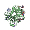
| Title | Crystal structure of the (BACCR)NAT3 arylamine N-acetyltransferase from Bacillus cereus reveals a unique Cys-His-Glu catalytic triad | ||||||
 Components Components | N-hydroxyarylamine O-acetyltransferase | ||||||
 Keywords Keywords | TRANSFERASE / Acetyltransferase | ||||||
| Function / homology |  Function and homology information Function and homology informationN-hydroxyarylamine O-acetyltransferase / N-hydroxyarylamine O-acetyltransferase activity Similarity search - Function | ||||||
| Biological species |  | ||||||
| Method |  X-RAY DIFFRACTION / SYNCHROTRON / MOLECULAR REPLACEMENT / Resolution: 2.14 Å X-RAY DIFFRACTION / SYNCHROTRON / MOLECULAR REPLACEMENT / Resolution: 2.14 Å | ||||||
 Authors Authors | Kubiak, X. / Li de la Sierra-Gallay, I. / Haouz, A. / Weber, P. / Rodrigues-Lima, F. | ||||||
 Citation Citation | Journal: J.Biol.Chem. / Year: 2013 Title: Structural and Biochemical Characterization of an Active Arylamine N-Acetyltransferase Possessing a Non-canonical Cys-His-Glu Catalytic Triad. Authors: Kubiak, X. / Li de la Sierra-Gallay, I. / Chaffotte, A.F. / Pluvinage, B. / Weber, P. / Haouz, A. / Dupret, J.M. / Rodrigues-Lima, F. | ||||||
| History |
|
- Structure visualization
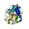
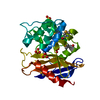
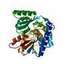
| Structure viewer | Molecule: MolmilJmol/JSmol |
|---|
- Downloads & links
-Download
| PDBx/mmCIF format | 4dmo.cif.gz | 199.7 KB | Display | PDBx/mmCIF format |
|---|---|---|---|---|
| PDB format | pdb4dmo.ent.gz | 163.4 KB | Display | PDB format |
| PDBx/mmJSON format | 4dmo.json.gz | Tree view | PDBx/mmJSON format | |
| Others |  Other downloads Other downloads |
-Validation report
| Arichive directory | https://data.pdbj.org/pub/pdb/validation_reports/dm/4dmoftp://data.pdbj.org/pub/pdb/validation_reports/dm/4dmo | HTTPS FTP |
|---|
-Related structure data
| Related structure data |  3lnbS S: Starting model for refinement |
|---|---|
| Similar structure data |


-Links
 PDBj
PDBj- Assembly
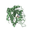
| Deposited unit | 
| ||||||||
|---|---|---|---|---|---|---|---|---|---|
| 1 | 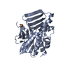
| ||||||||
| 2 | 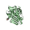
| ||||||||
| Unit cell |
|
-Components
| #1: Protein | Mass: 30100.010 Da / Num. of mol.: 2 Source method: isolated from a genetically manipulated source Source: (gene. exp.) References: UniProt: Q81AS3, N-hydroxyarylamine O-acetyltransferase #2: Chemical | 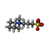  Mass: 221.317 Da / Num. of mol.: 3 / Source method: obtained synthetically / Formula: C9H19NO3S Mass: 221.317 Da / Num. of mol.: 3 / Source method: obtained synthetically / Formula: C9H19NO3S#3: Water | ChemComp-HOH / |  Mass: 18.015 Da / Num. of mol.: 128 / Source method: isolated from a natural source / Formula: H2O Mass: 18.015 Da / Num. of mol.: 128 / Source method: isolated from a natural source / Formula: H2O |
|---|
-Experimental details
-Experiment
| Experiment | Method: X-RAY DIFFRACTION / Number of used crystals: 1 |
|---|
- Sample preparation
| Crystal | Density Matthews: 2.16 Å3/Da / Density % sol: 43.03 % |
|---|---|
| Crystal grow | Temperature: 291 K / Method: vapor diffusion, hanging drop / pH: 6.5 Details: 1.37 M sodium citrate, 0.28 M NDSB-221, pH 6.5, VAPOR DIFFUSION, HANGING DROP, temperature 291K |
-Data collection
| Diffraction | Mean temperature: 100 K |
|---|---|
| Diffraction source | Source: SYNCHROTRON / Site: SLS  / Beamline: X06DA / Wavelength: 0.984 Å / Beamline: X06DA / Wavelength: 0.984 Å |
| Detector | Type: MARMOSAIC 225 mm CCD / Detector: CCD / Date: Aug 20, 2011 / Details: collimating (M1) and toroidal (M2) mirrors |
| Radiation | Monochromator: Bartels Monochromator / Protocol: SINGLE WAVELENGTH / Monochromatic (M) / Laue (L): M / Scattering type: x-ray |
| Radiation wavelength | Wavelength: 0.984 Å / Relative weight: 1 |
| Reflection | Resolution: 2.14→19.967 Å / Num. obs: 28380 / % possible obs: 99.3 % / Observed criterion σ(I): -3 / Redundancy: 7.392 % / Biso Wilson estimate: 45.83 Å2 / Rmerge(I) obs: 0.113 / Net I/σ(I): 19.07 |
| Reflection shell | Resolution: 2.14→2.22 Å / Redundancy: 7.25 % / Rmerge(I) obs: 0.437 / Mean I/σ(I) obs: 4.28 / % possible all: 97.2 |
- Processing
| Software |
| |||||||||||||||||||||||||||||||||||||||||||||||||||||||||||||||||||||||||||||
|---|---|---|---|---|---|---|---|---|---|---|---|---|---|---|---|---|---|---|---|---|---|---|---|---|---|---|---|---|---|---|---|---|---|---|---|---|---|---|---|---|---|---|---|---|---|---|---|---|---|---|---|---|---|---|---|---|---|---|---|---|---|---|---|---|---|---|---|---|---|---|---|---|---|---|---|---|---|---|
| Refinement | Method to determine structure: MOLECULAR REPLACEMENT Starting model: PDB ENTRY 3LNB Resolution: 2.14→19.95 Å / Cor.coef. Fo:Fc: 0.9417 / Cor.coef. Fo:Fc free: 0.9062 / SU ML: 0.28 / SU R Cruickshank DPI: 0.593 / σ(F): 1.99 / Phase error: 31.95 / Stereochemistry target values: ML Details: Additional restraint dictionnary file used through the Gelly module implemented in Buster: protgeo_option_chiralrestraint_from_equilib.dat
| |||||||||||||||||||||||||||||||||||||||||||||||||||||||||||||||||||||||||||||
| Solvent computation | Shrinkage radii: 0.9 Å / VDW probe radii: 1.11 Å / Solvent model: FLAT BULK SOLVENT MODEL | |||||||||||||||||||||||||||||||||||||||||||||||||||||||||||||||||||||||||||||
| Displacement parameters | Biso mean: 37.81 Å2
| |||||||||||||||||||||||||||||||||||||||||||||||||||||||||||||||||||||||||||||
| Refine analyze | Luzzati coordinate error obs: 0.341 Å | |||||||||||||||||||||||||||||||||||||||||||||||||||||||||||||||||||||||||||||
| Refinement step | Cycle: LAST / Resolution: 2.14→19.95 Å
| |||||||||||||||||||||||||||||||||||||||||||||||||||||||||||||||||||||||||||||
| Refine LS restraints |
| |||||||||||||||||||||||||||||||||||||||||||||||||||||||||||||||||||||||||||||
| LS refinement shell |
|
