+ Open data
- Basic information
| Entry | Database: PDB / ID: 6nwq | |||||||||||||||||||||
|---|---|---|---|---|---|---|---|---|---|---|---|---|---|---|---|---|---|---|---|---|---|---|
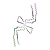
| Title | Chronic traumatic encephalopathy Type II Tau filament | |||||||||||||||||||||
 Components Components | Microtubule-associated protein tau | |||||||||||||||||||||
 Keywords Keywords | PROTEIN FIBRIL / Tau / chronic traumatic encephalopathy / filament / amyloid | |||||||||||||||||||||
| Function / homology |  Function and homology information Function and homology informationplus-end-directed organelle transport along microtubule / histone-dependent DNA binding / negative regulation of protein localization to mitochondrion / neurofibrillary tangle / microtubule lateral binding / axonal transport / tubulin complex / positive regulation of protein localization to synapse / phosphatidylinositol bisphosphate binding / generation of neurons ...plus-end-directed organelle transport along microtubule / histone-dependent DNA binding / negative regulation of protein localization to mitochondrion / neurofibrillary tangle / microtubule lateral binding / axonal transport / tubulin complex / positive regulation of protein localization to synapse / phosphatidylinositol bisphosphate binding / generation of neurons / rRNA metabolic process / axonal transport of mitochondrion / regulation of mitochondrial fission / axon development / regulation of chromosome organization / regulation of microtubule-based movement / central nervous system neuron development / intracellular distribution of mitochondria / minor groove of adenine-thymine-rich DNA binding / lipoprotein particle binding / microtubule polymerization / negative regulation of mitochondrial membrane potential / regulation of microtubule polymerization / dynactin binding / apolipoprotein binding / main axon / protein polymerization / axolemma / glial cell projection / Caspase-mediated cleavage of cytoskeletal proteins / regulation of microtubule polymerization or depolymerization / negative regulation of mitochondrial fission / neurofibrillary tangle assembly / positive regulation of axon extension / regulation of cellular response to heat / synapse assembly / Activation of AMPK downstream of NMDARs / regulation of long-term synaptic depression / positive regulation of superoxide anion generation / positive regulation of protein localization / cellular response to brain-derived neurotrophic factor stimulus / supramolecular fiber organization / cytoplasmic microtubule organization / regulation of calcium-mediated signaling / somatodendritic compartment / axon cytoplasm / positive regulation of microtubule polymerization / astrocyte activation / phosphatidylinositol binding / enzyme inhibitor activity / nuclear periphery / stress granule assembly / protein phosphatase 2A binding / regulation of microtubule cytoskeleton organization / cellular response to reactive oxygen species / Hsp90 protein binding / microglial cell activation / cellular response to nerve growth factor stimulus / synapse organization / PKR-mediated signaling / regulation of synaptic plasticity / protein homooligomerization / response to lead ion / SH3 domain binding / microtubule cytoskeleton organization / memory / regulation of autophagy / cytoplasmic ribonucleoprotein granule / neuron projection development / cell-cell signaling / single-stranded DNA binding / protein-folding chaperone binding / cellular response to heat / microtubule cytoskeleton / growth cone / actin binding / cell body / double-stranded DNA binding / protein-macromolecule adaptor activity / microtubule binding / sequence-specific DNA binding / dendritic spine / amyloid fibril formation / microtubule / learning or memory / neuron projection / membrane raft / axon / negative regulation of gene expression / neuronal cell body / DNA damage response / dendrite / protein kinase binding / enzyme binding / mitochondrion / DNA binding / RNA binding / extracellular region / identical protein binding / nucleus Similarity search - Function | |||||||||||||||||||||
| Biological species |  Homo sapiens (human) Homo sapiens (human) | |||||||||||||||||||||
| Method | ELECTRON MICROSCOPY / helical reconstruction / cryo EM / Resolution: 3.4 Å | |||||||||||||||||||||
 Authors Authors | Falcon, B. / Zivanov, J. / Zhang, W. / Murzin, A.G. / Garringer, H.J. / Vidal, R. / Crowther, R.A. / Newell, K.L. / Ghetti, B. / Goedert, M. / Scheres, H.W. | |||||||||||||||||||||
| Funding support |  United Kingdom, United Kingdom,  United States, 6items United States, 6items
| |||||||||||||||||||||
 Citation Citation |  Journal: Nature / Year: 2019 Journal: Nature / Year: 2019Title: Novel tau filament fold in chronic traumatic encephalopathy encloses hydrophobic molecules. Authors: Benjamin Falcon / Jasenko Zivanov / Wenjuan Zhang / Alexey G Murzin / Holly J Garringer / Ruben Vidal / R Anthony Crowther / Kathy L Newell / Bernardino Ghetti / Michel Goedert / Sjors H W Scheres /   Abstract: Chronic traumatic encephalopathy (CTE) is a neurodegenerative tauopathy that is associated with repetitive head impacts or exposure to blast waves. First described as punch-drunk syndrome and ...Chronic traumatic encephalopathy (CTE) is a neurodegenerative tauopathy that is associated with repetitive head impacts or exposure to blast waves. First described as punch-drunk syndrome and dementia pugilistica in retired boxers, CTE has since been identified in former participants of other contact sports, ex-military personnel and after physical abuse. No disease-modifying therapies currently exist, and diagnosis requires an autopsy. CTE is defined by an abundance of hyperphosphorylated tau protein in neurons, astrocytes and cell processes around blood vessels. This, together with the accumulation of tau inclusions in cortical layers II and III, distinguishes CTE from Alzheimer's disease and other tauopathies. However, the morphologies of tau filaments in CTE and the mechanisms by which brain trauma can lead to their formation are unknown. Here we determine the structures of tau filaments from the brains of three individuals with CTE at resolutions down to 2.3 Å, using cryo-electron microscopy. We show that filament structures are identical in the three cases but are distinct from those of Alzheimer's and Pick's diseases, and from those formed in vitro. Similar to Alzheimer's disease, all six brain tau isoforms assemble into filaments in CTE, and residues K274-R379 of three-repeat tau and S305-R379 of four-repeat tau form the ordered core of two identical C-shaped protofilaments. However, a different conformation of the β-helix region creates a hydrophobic cavity that is absent in tau filaments from the brains of patients with Alzheimer's disease. This cavity encloses an additional density that is not connected to tau, which suggests that the incorporation of cofactors may have a role in tau aggregation in CTE. Moreover, filaments in CTE have distinct protofilament interfaces to those of Alzheimer's disease. Our structures provide a unifying neuropathological criterion for CTE, and support the hypothesis that the formation and propagation of distinct conformers of assembled tau underlie different neurodegenerative diseases. | |||||||||||||||||||||
| History |
|
- Structure visualization
| Movie |
 Movie viewer Movie viewer |
|---|---|
| Structure viewer | Molecule:  Molmil Molmil Jmol/JSmol Jmol/JSmol |
- Downloads & links
- Download
| PDBx/mmCIF format |  6nwq.cif.gz 6nwq.cif.gz | 123.8 KB | Display |  PDBx/mmCIF format PDBx/mmCIF format |
|---|---|---|---|---|
| PDB format |  pdb6nwq.ent.gz pdb6nwq.ent.gz | 82 KB | Display |  PDB format PDB format |
| PDBx/mmJSON format |  6nwq.json.gz 6nwq.json.gz | Tree view |  PDBx/mmJSON format PDBx/mmJSON format | |
| Others |  Other downloads Other downloads |
-Validation report
| Arichive directory |  https://data.pdbj.org/pub/pdb/validation_reports/nw/6nwq https://data.pdbj.org/pub/pdb/validation_reports/nw/6nwq ftp://data.pdbj.org/pub/pdb/validation_reports/nw/6nwq ftp://data.pdbj.org/pub/pdb/validation_reports/nw/6nwq | HTTPS FTP |
|---|
-Related structure data
| Related structure data |  0528MC  0527C  6nwpC C: citing same article ( M: map data used to model this data |
|---|---|
| Similar structure data | |
| EM raw data |  EMPIAR-10313 (Title: Cryo-EM reconstructions of tau filaments in chronic traumatic encephalopathy EMPIAR-10313 (Title: Cryo-EM reconstructions of tau filaments in chronic traumatic encephalopathyData size: 9.7 TB / Data #1: Raw Movies [micrographs - multiframe] / Data #2: Aligned Micrographs [micrographs - single frame]) |
- Links
Links
- Assembly
| Deposited unit | 
|
|---|---|
| 1 |
|
- Components
| #1: Protein | Mass: 45919.871 Da / Num. of mol.: 6 / Source method: isolated from a natural source / Source: (natural)  Homo sapiens (human) / Organ: Brain / Tissue: Temporal cortex / References: UniProt: P10636 Homo sapiens (human) / Organ: Brain / Tissue: Temporal cortex / References: UniProt: P10636 |
|---|
-Experimental details
-Experiment
| Experiment | Method: ELECTRON MICROSCOPY |
|---|---|
| EM experiment | Aggregation state: TISSUE / 3D reconstruction method: helical reconstruction |
- Sample preparation
| Component | Name: Tau filaments extracted from the temporal cortex of a patient with chronic traumatic encephalopathy Type: TISSUE / Entity ID: all / Source: NATURAL | ||||||||||||
|---|---|---|---|---|---|---|---|---|---|---|---|---|---|
| Source (natural) | Organism:  Homo sapiens (human) / Organ: Brain / Tissue: Temporal cortex Homo sapiens (human) / Organ: Brain / Tissue: Temporal cortex | ||||||||||||
| Buffer solution | pH: 7.4 | ||||||||||||
| Buffer component |
| ||||||||||||
| Specimen | Conc.: 0.5 mg/ml / Embedding applied: NO / Shadowing applied: NO / Staining applied: NO / Vitrification applied: YES | ||||||||||||
| Vitrification | Instrument: FEI VITROBOT MARK IV / Cryogen name: ETHANE / Humidity: 100 % / Chamber temperature: 277.15 K |
- Electron microscopy imaging
| Experimental equipment |  Model: Titan Krios / Image courtesy: FEI Company |
|---|---|
| Microscopy | Model: FEI TITAN KRIOS |
| Electron gun | Electron source:  FIELD EMISSION GUN / Accelerating voltage: 300 kV / Illumination mode: FLOOD BEAM FIELD EMISSION GUN / Accelerating voltage: 300 kV / Illumination mode: FLOOD BEAM |
| Electron lens | Mode: BRIGHT FIELD / Nominal magnification: 130000 X / Nominal defocus max: 2800 nm / Nominal defocus min: 1700 nm / Cs: 2.7 mm |
| Image recording | Average exposure time: 9 sec. / Electron dose: 55.48 e/Å2 / Detector mode: COUNTING / Film or detector model: GATAN K2 SUMMIT (4k x 4k) |
| EM imaging optics | Energyfilter name: GIF Quantum LS / Energyfilter slit width: 25 eV |
- Processing
| EM software |
| ||||||||||||||||||||
|---|---|---|---|---|---|---|---|---|---|---|---|---|---|---|---|---|---|---|---|---|---|
| CTF correction | Type: PHASE FLIPPING AND AMPLITUDE CORRECTION | ||||||||||||||||||||
| Helical symmerty | Angular rotation/subunit: 179.4 ° / Axial rise/subunit: 2.37 Å / Axial symmetry: C2 | ||||||||||||||||||||
| 3D reconstruction | Resolution: 3.4 Å / Resolution method: FSC 0.143 CUT-OFF / Num. of particles: 7621 / Symmetry type: HELICAL | ||||||||||||||||||||
| Atomic model building | B value: 51.7 / Space: RECIPROCAL / Target criteria: Fourier shell correlation Details: Fourier-space refinement of the complete atomic model against the chronic traumatic encephalopathy Type II Tau filament map was performed in REFMAC. A stack of three consecutive monomers was ...Details: Fourier-space refinement of the complete atomic model against the chronic traumatic encephalopathy Type II Tau filament map was performed in REFMAC. A stack of three consecutive monomers was refined to preserve nearest-neighbour interactions for the middle chain. |
 Movie
Movie Controller


 PDBj
PDBj


