 Movie
Movie Controller


+ Open data

- Basic information
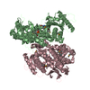
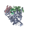
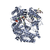
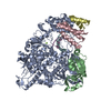
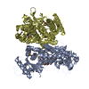
| Entry | Database: PDB / ID: 6nus | |||||||||
|---|---|---|---|---|---|---|---|---|---|---|
| Title | SARS-Coronavirus NSP12 bound to NSP8 co-factor | |||||||||
 Components Components |
| |||||||||
 Keywords Keywords | VIRAL PROTEIN / coronavirus / polymerase / non-structural protein | |||||||||
| Function / homology |  Function and homology information Function and homology informationAssembly of the SARS-CoV-1 Replication-Transcription Complex (RTC) / Maturation of replicase proteins / Transcription of SARS-CoV-1 sgRNAs / Translation of Replicase and Assembly of the Replication Transcription Complex / K48-linked deubiquitinase activity / Replication of the SARS-CoV-1 genome / K63-linked deubiquitinase activity / host cell endoplasmic reticulum / viral transcription / SARS-CoV-1 modulates host translation machinery ...Assembly of the SARS-CoV-1 Replication-Transcription Complex (RTC) / Maturation of replicase proteins / Transcription of SARS-CoV-1 sgRNAs / Translation of Replicase and Assembly of the Replication Transcription Complex / K48-linked deubiquitinase activity / Replication of the SARS-CoV-1 genome / K63-linked deubiquitinase activity / host cell endoplasmic reticulum / viral transcription / SARS-CoV-1 modulates host translation machinery / Transferases; Transferring one-carbon groups; Methyltransferases / viral genome replication / methyltransferase activity / SARS-CoV-1 activates/modulates innate immune responses / 5'-3' RNA helicase activity / endonuclease activity / Lyases; Phosphorus-oxygen lyases / methylation / Hydrolases; Acting on ester bonds; Exoribonucleases producing 5'-phosphomonoesters / double membrane vesicle viral factory outer membrane / SARS coronavirus main proteinase / host cell endoplasmic reticulum-Golgi intermediate compartment / host cell endosome / 3'-5'-RNA exonuclease activity / symbiont-mediated degradation of host mRNA / 5'-3' DNA helicase activity / mRNA guanylyltransferase / symbiont-mediated suppression of host ISG15-protein conjugation / G-quadruplex RNA binding / mRNA guanylyltransferase activity / symbiont-mediated suppression of host cytoplasmic pattern recognition receptor signaling pathway via inhibition of IRF3 activity / omega peptidase activity / mRNA (guanine-N7)-methyltransferase / methyltransferase cap1 / DNA helicase / symbiont-mediated suppression of host NF-kappaB cascade / symbiont-mediated perturbation of host ubiquitin-like protein modification / host cell Golgi apparatus / methyltransferase cap1 activity / mRNA 5'-cap (guanine-N7-)-methyltransferase activity / cysteine-type deubiquitinase activity / ubiquitinyl hydrolase 1 / Hydrolases; Acting on peptide bonds (peptidases); Cysteine endopeptidases / lyase activity / single-stranded RNA binding / viral protein processing / host cell perinuclear region of cytoplasm / RNA helicase / symbiont-mediated suppression of host type I interferon-mediated signaling pathway / symbiont-mediated suppression of host gene expression / viral translational frameshifting / symbiont-mediated activation of host autophagy / RNA-directed RNA polymerase / cysteine-type endopeptidase activity / viral RNA genome replication / RNA-directed RNA polymerase activity / DNA-templated transcription / ATP hydrolysis activity / proteolysis / zinc ion binding / ATP binding / identical protein binding Similarity search - Function | |||||||||
| Biological species |   Human SARS coronavirus Human SARS coronavirus | |||||||||
| Method | ELECTRON MICROSCOPY / single particle reconstruction / cryo EM / Resolution: 3.5 Å | |||||||||
 Authors Authors | Kirchdoerfer, R.N. / Ward, A.B. | |||||||||
| Funding support |  United States, 2items United States, 2items
| |||||||||
 Citation Citation | Journal: Nat Commun / Year: 2019 Title: Structure of the SARS-CoV nsp12 polymerase bound to nsp7 and nsp8 co-factors. Authors: Robert N Kirchdoerfer / Andrew B Ward / Abstract: Recent history is punctuated by the emergence of highly pathogenic coronaviruses such as SARS- and MERS-CoV into human circulation. Upon infecting host cells, coronaviruses assemble a multi-subunit ...Recent history is punctuated by the emergence of highly pathogenic coronaviruses such as SARS- and MERS-CoV into human circulation. Upon infecting host cells, coronaviruses assemble a multi-subunit RNA-synthesis complex of viral non-structural proteins (nsp) responsible for the replication and transcription of the viral genome. Here, we present the 3.1 Å resolution structure of the SARS-CoV nsp12 polymerase bound to its essential co-factors, nsp7 and nsp8, using single particle cryo-electron microscopy. nsp12 possesses an architecture common to all viral polymerases as well as a large N-terminal extension containing a kinase-like fold and is bound by two nsp8 co-factors. This structure illuminates the assembly of the coronavirus core RNA-synthesis machinery, provides key insights into nsp12 polymerase catalysis and fidelity and acts as a template for the design of novel antiviral therapeutics. | |||||||||
| History |
|
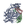
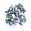
- Structure visualization
| Movie |
Movie viewer |
|---|---|
| Structure viewer | Molecule: MolmilJmol/JSmol |
- Downloads & links
-Download
| PDBx/mmCIF format | 6nus.cif.gz | 181 KB | Display | PDBx/mmCIF format |
|---|---|---|---|---|
| PDB format | pdb6nus.ent.gz | 136.7 KB | Display | PDB format |
| PDBx/mmJSON format | 6nus.json.gz | Tree view | PDBx/mmJSON format | |
| Others |  Other downloads Other downloads |
-Validation report
| Arichive directory | https://data.pdbj.org/pub/pdb/validation_reports/nu/6nusftp://data.pdbj.org/pub/pdb/validation_reports/nu/6nus | HTTPS FTP |
|---|
-Related structure data
| Related structure data | 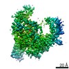 0521MC 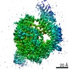 0520C 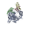 6nurC M: map data used to model this data C: citing same article ( |
|---|---|
| Similar structure data |
-Links
 PDBj
PDBj


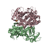
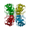
- Assembly
| Deposited unit | 
|
|---|---|
| 1 |
|
-Components
| #1: Protein | Mass: 109277.031 Da / Num. of mol.: 1 / Fragment: UNP residues 4370-5300 Source method: isolated from a genetically manipulated source Source: (gene. exp.) Human SARS coronavirus / Gene: rep, 1a-1b / Plasmid: pFastBac / Cell line (production host): Sf21 / Production host:   Spodoptera frugiperda (fall armyworm) / References: UniProt: P0C6X7 Spodoptera frugiperda (fall armyworm) / References: UniProt: P0C6X7 |
|---|---|
| #2: Protein | Mass: 21887.990 Da / Num. of mol.: 1 / Fragment: UNP residues 3920-4117 Source method: isolated from a genetically manipulated source Source: (gene. exp.) Human SARS coronavirus / Gene: 1a / Production host:  |
| #3: Chemical |   Mass: 65.409 Da / Num. of mol.: 2 / Source method: obtained synthetically / Formula: Zn Mass: 65.409 Da / Num. of mol.: 2 / Source method: obtained synthetically / Formula: Zn |
-Experimental details
-Experiment
| Experiment | Method: ELECTRON MICROSCOPY |
|---|---|
| EM experiment | Aggregation state: PARTICLE / 3D reconstruction method: single particle reconstruction |
- Sample preparation
| Component | Name: SARS-Coronavirus NSP12 bound to NSP7 and NSP8 co-factors Type: COMPLEX / Entity ID: #1-#2 / Source: RECOMBINANT | ||||||||||||||||||||||||||||||
|---|---|---|---|---|---|---|---|---|---|---|---|---|---|---|---|---|---|---|---|---|---|---|---|---|---|---|---|---|---|---|---|
| Molecular weight | Value: 0.13 MDa / Experimental value: NO | ||||||||||||||||||||||||||||||
| Source (natural) | Organism: SARS coronavirus | ||||||||||||||||||||||||||||||
| Source (recombinant) | Organism: Spodoptera frugiperda (fall armyworm) / Cell: Sf21 / Plasmid: pFastBac | ||||||||||||||||||||||||||||||
| Buffer solution | pH: 7.4 Details: n-dodecyl-beta-D-maltopyranoside was added just prior to spotting samples onto holey EM grids. | ||||||||||||||||||||||||||||||
| Buffer component |
| ||||||||||||||||||||||||||||||
| Specimen | Conc.: 3.1 mg/ml / Embedding applied: NO / Shadowing applied: NO / Staining applied: NO / Vitrification applied: YES | ||||||||||||||||||||||||||||||
| Specimen support | Grid material: GOLD / Grid mesh size: 300 divisions/in. / Grid type: Quantifoil, UltrAuFoil, R1.2/1.3 | ||||||||||||||||||||||||||||||
| Vitrification | Instrument: FEI VITROBOT MARK IV / Cryogen name: ETHANE / Humidity: 100 % / Chamber temperature: 277 K |
- Electron microscopy imaging
| Experimental equipment |  Model: Talos Arctica / Image courtesy: FEI Company |
|---|---|
| Microscopy | Model: FEI TALOS ARCTICA |
| Electron gun | Electron source:  FIELD EMISSION GUN / Accelerating voltage: 200 kV / Illumination mode: FLOOD BEAM FIELD EMISSION GUN / Accelerating voltage: 200 kV / Illumination mode: FLOOD BEAM |
| Electron lens | Mode: BRIGHT FIELD / Calibrated magnification: 43478 X / Calibrated defocus min: 400 nm / Calibrated defocus max: 1000 nm / Cs: 2.7 mm / C2 aperture diameter: 70 µm |
| Specimen holder | Cryogen: NITROGEN / Specimen holder model: FEI TITAN KRIOS AUTOGRID HOLDER |
| Image recording | Average exposure time: 11.75 sec. / Electron dose: 50.5 e/Å2 / Detector mode: COUNTING / Film or detector model: GATAN K2 SUMMIT (4k x 4k) / Num. of grids imaged: 1 / Num. of real images: 1677 |
| Image scans | Width: 3838 / Height: 3710 / Movie frames/image: 47 / Used frames/image: 1-47 |
- Processing
| EM software |
| ||||||||||||||||||||||||||||||||||||||||||||
|---|---|---|---|---|---|---|---|---|---|---|---|---|---|---|---|---|---|---|---|---|---|---|---|---|---|---|---|---|---|---|---|---|---|---|---|---|---|---|---|---|---|---|---|---|---|
| CTF correction | Type: PHASE FLIPPING AND AMPLITUDE CORRECTION | ||||||||||||||||||||||||||||||||||||||||||||
| Particle selection | Num. of particles selected: 2003890 | ||||||||||||||||||||||||||||||||||||||||||||
| Symmetry | Point symmetry: C1 (asymmetric) | ||||||||||||||||||||||||||||||||||||||||||||
| 3D reconstruction | Resolution: 3.5 Å / Resolution method: FSC 0.143 CUT-OFF / Num. of particles: 71262 / Symmetry type: POINT | ||||||||||||||||||||||||||||||||||||||||||||
| Atomic model building | Space: REAL |
