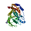
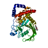
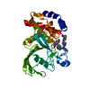
Tyrosine-protein phosphatase non-receptor type 1,NPH1-1
Keywords
HYDROLASE / Protein tyrosine phosphatase / protein phosphorylation / signaling / diabetes / obesity / cancer
Function / homology
Function and homology information
blue light photoreceptor activity / PTK6 Down-Regulation / regulation of hepatocyte growth factor receptor signaling pathway / positive regulation of receptor catabolic process / insulin receptor recycling / negative regulation of vascular endothelial growth factor receptor signaling pathway / regulation of intracellular protein transport / negative regulation of MAP kinase activity / mitochondrial crista / IRE1-mediated unfolded protein response ...blue light photoreceptor activity / PTK6 Down-Regulation / regulation of hepatocyte growth factor receptor signaling pathway / positive regulation of receptor catabolic process / insulin receptor recycling / negative regulation of vascular endothelial growth factor receptor signaling pathway / regulation of intracellular protein transport / negative regulation of MAP kinase activity / mitochondrial crista / IRE1-mediated unfolded protein response / platelet-derived growth factor receptor-beta signaling pathway / sorting endosome / positive regulation of IRE1-mediated unfolded protein response / negative regulation of PERK-mediated unfolded protein response / regulation of type I interferon-mediated signaling pathway / cytoplasmic side of endoplasmic reticulum membrane / negative regulation of vascular associated smooth muscle cell migration / vascular endothelial cell response to oscillatory fluid shear stress / peptidyl-tyrosine dephosphorylation / positive regulation of systemic arterial blood pressure / non-membrane spanning protein tyrosine phosphatase activity / Regulation of IFNA/IFNB signaling / regulation of endocytosis / cellular response to angiotensin / regulation of proteolysis / growth hormone receptor signaling pathway via JAK-STAT / negative regulation of cell-substrate adhesion / cellular response to unfolded protein / regulation of postsynapse assembly / positive regulation of endothelial cell apoptotic process / regulation of signal transduction / negative regulation of signal transduction / Regulation of IFNG signaling / Growth hormone receptor signaling / negative regulation of endoplasmic reticulum stress-induced intrinsic apoptotic signaling pathway / positive regulation of heart rate / ephrin receptor binding / cellular response to platelet-derived growth factor stimulus / Insulin receptor recycling / MECP2 regulates neuronal receptors and channels / Integrin signaling / endoplasmic reticulum unfolded protein response / phosphoprotein phosphatase activity / protein-tyrosine-phosphatase / cellular response to fibroblast growth factor stimulus / cellular response to nitric oxide / negative regulation of insulin receptor signaling pathway / positive regulation of cardiac muscle cell apoptotic process / protein tyrosine phosphatase activity / protein phosphatase 2A binding / Turbulent (oscillatory, disturbed) flow shear stress activates signaling by PIEZO1 and integrins in endothelial cells / endosome lumen / negative regulation of phosphatidylinositol 3-kinase/protein kinase B signal transduction / insulin receptor binding / cellular response to nerve growth factor stimulus / negative regulation of ERK1 and ERK2 cascade / response to nutrient levels / Negative regulation of MET activity / receptor tyrosine kinase binding / positive regulation of JNK cascade / insulin receptor signaling pathway / negative regulation of neuron projection development / actin cytoskeleton organization / cellular response to hypoxia / early endosome / non-specific serine/threonine protein kinase / postsynapse / cadherin binding / mitochondrial matrix / negative regulation of cell population proliferation / protein serine/threonine kinase activity / protein kinase binding / glutamatergic synapse / enzyme binding / endoplasmic reticulum / protein-containing complex / RNA binding / zinc ion binding / ATP binding / metal ion binding / cytosol / cytoplasm Similarity search - Function
PAS domain / PAS-associated, C-terminal / PAC domain profile. / PAC motif / Motif C-terminal to PAS motifs (likely to contribute to PAS structural domain) / Protein-tyrosine phosphatase, non-receptor type-1/2 / : / Protein tyrosine phosphatase superfamily / Protein-Tyrosine Phosphatase; Chain A / Protein tyrosine phosphatase, catalytic domain ...PAS domain / PAS-associated, C-terminal / PAC domain profile. / PAC motif / Motif C-terminal to PAS motifs (likely to contribute to PAS structural domain) / Protein-tyrosine phosphatase, non-receptor type-1/2 / : / Protein tyrosine phosphatase superfamily / Protein-Tyrosine Phosphatase; Chain A / Protein tyrosine phosphatase, catalytic domain / PTP type protein phosphatase domain profile. / Protein-tyrosine phosphatase / Tyrosine-specific protein phosphatase, PTPase domain / Protein-tyrosine phosphatase, catalytic / Protein tyrosine phosphatase, catalytic domain motif / Tyrosine specific protein phosphatases active site. / Protein-tyrosine phosphatase, active site / Tyrosine specific protein phosphatases domain profile. / Tyrosine-specific protein phosphatases domain / Protein-tyrosine phosphatase-like / PAS domain / PAS repeat profile. / PAS domain / PAS domain superfamily / Serine/threonine-protein kinase, active site / Serine/Threonine protein kinases active-site signature. / Protein kinase domain / Serine/Threonine protein kinases, catalytic domain / Protein kinase, ATP binding site / Protein kinases ATP-binding region signature. / Protein kinase domain profile. / Protein kinase domain / Alpha-Beta Complex / Protein kinase-like domain superfamily / Alpha Beta Similarity search - Domain/homology
non-specific serine/threonine protein kinase / Tyrosine-protein phosphatase non-receptor type 1 Similarity search - Component
Resolution: 1.89→44.67 Å / Cor.coef. Fo:Fc: 0.97 / Cor.coef. Fo:Fc free: 0.954 / SU B: 6.223 / SU ML: 0.087 / Cross valid method: THROUGHOUT / ESU R: 0.107 / ESU R Free: 0.107 / Stereochemistry target values: MAXIMUM LIKELIHOOD / Details: HYDROGENS HAVE BEEN ADDED IN THE RIDING POSITIONS
Rfactor
Num. reflection
% reflection
Selection details
Rfree
0.21175
1947
5 %
RANDOM
Rwork
0.17922
-
-
-
obs
0.18073
37270
99.27 %
-
Solvent computation
Ion probe radii: 0.7 Å / Shrinkage radii: 0.7 Å / VDW probe radii: 1 Å / Solvent model: MASK
 Movie
Movie Controller


 Open data
 Basic information
 Components
 Keywords
 Function and homology information
 Homo sapiens (human)
 X-RAY DIFFRACTION /
  Authors
 United States, 3items
  Citation
 Structure visualization
 Downloads & links
 Other downloads
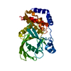


 PDBj
PDBj


 Assembly
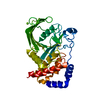


 Mass: 24.305 Da / Num. of mol.: 1 / Source method: obtained synthetically / Formula: Mg
 Mass: 18.015 Da / Num. of mol.: 224 / Source method: isolated from a natural source / Formula: H2O
 Sample preparation
 Processing