lysine-tRNA ligase / lysine-tRNA ligase activity / lysyl-tRNA aminoacylation / tRNA binding / magnesium ion binding / ATP binding / cytosol Similarity search - Function
Lysine-tRNA ligase, class II / Lysine-tRNA ligase, class II, N-terminal / Lysyl-tRNA synthetase, class II, C-terminal / Aminoacyl-tRNA synthetase, class II (D/K/N) / tRNA synthetases class II (D, K and N) / OB-fold nucleic acid binding domain, AA-tRNA synthetase-type / OB-fold nucleic acid binding domain / Bira Bifunctional Protein; Domain 2 / BirA Bifunctional Protein; domain 2 / Aminoacyl-tRNA synthetase, class II ...Lysine-tRNA ligase, class II / Lysine-tRNA ligase, class II, N-terminal / Lysyl-tRNA synthetase, class II, C-terminal / Aminoacyl-tRNA synthetase, class II (D/K/N) / tRNA synthetases class II (D, K and N) / OB-fold nucleic acid binding domain, AA-tRNA synthetase-type / OB-fold nucleic acid binding domain / Bira Bifunctional Protein; Domain 2 / BirA Bifunctional Protein; domain 2 / Aminoacyl-tRNA synthetase, class II / Aminoacyl-transfer RNA synthetases class-II family profile. / Class II Aminoacyl-tRNA synthetase/Biotinyl protein ligase (BPL) and lipoyl protein ligase (LPL) / Nucleic acid-binding proteins / OB fold (Dihydrolipoamide Acetyltransferase, E2P) / Nucleic acid-binding, OB-fold / Beta Barrel / 2-Layer Sandwich / Mainly Beta / Alpha Beta Similarity search - Domain/homology
Mass: 18.015 Da / Num. of mol.: 463 / Source method: isolated from a natural source / Formula: H2O
-
Details
Has protein modification
N
-
Experimental details
-
Experiment
Experiment
Method: X-RAY DIFFRACTION / Number of used crystals: 1
-
Sample preparation
Crystal
Density Matthews: 3.14 Å3/Da / Density % sol: 60.87 %
Crystal grow
Temperature: 287 K / Method: vapor diffusion, sitting drop / pH: 7.5 Details: ChtrA.00612.a.B1.PW38393 at 25.76 mg/ml was incubated with 3 mM l-lysine, 3 mM cladosporin, then was mixed 1:1 MorpheusB6opt1(e11): 10.96% (w/v) PEG 8000, 21.92% (v/v) ethylene glycol, 0.03 ...Details: ChtrA.00612.a.B1.PW38393 at 25.76 mg/ml was incubated with 3 mM l-lysine, 3 mM cladosporin, then was mixed 1:1 MorpheusB6opt1(e11): 10.96% (w/v) PEG 8000, 21.92% (v/v) ethylene glycol, 0.03 M each sodium fluoride, sodium bromide, sodium iodide, 0.1 M MOPE/HEPES-Na, pH=7.5. Tray: 300519e11, puck: ytb8-10. Crystallization of protein from strain UCH-1/proctitis and strain D/UW-3/Cx gave the same data.
-
Data collection
Diffraction
Mean temperature: 100 K / Serial crystal experiment: N
In the structure databanks used in Yorodumi, some data are registered as the other names, "COVID-19 virus" and "2019-nCoV". Here are the details of the virus and the list of structure data.
Jan 31, 2019. EMDB accession codes are about to change! (news from PDBe EMDB page)
EMDB accession codes are about to change! (news from PDBe EMDB page)
The allocation of 4 digits for EMDB accession codes will soon come to an end. Whilst these codes will remain in use, new EMDB accession codes will include an additional digit and will expand incrementally as the available range of codes is exhausted. The current 4-digit format prefixed with “EMD-” (i.e. EMD-XXXX) will advance to a 5-digit format (i.e. EMD-XXXXX), and so on. It is currently estimated that the 4-digit codes will be depleted around Spring 2019, at which point the 5-digit format will come into force.
The EM Navigator/Yorodumi systems omit the EMD- prefix.
Related info.:Q: What is EMD? / ID/Accession-code notation in Yorodumi/EM Navigator
Yorodumi is a browser for structure data from EMDB, PDB, SASBDB, etc.
This page is also the successor to EM Navigator detail page, and also detail information page/front-end page for Omokage search.
The word "yorodu" (or yorozu) is an old Japanese word meaning "ten thousand". "mi" (miru) is to see.
Related info.:EMDB / PDB / SASBDB / Comparison of 3 databanks / Yorodumi Search / Aug 31, 2016. New EM Navigator & Yorodumi / Yorodumi Papers / Jmol/JSmol / Function and homology information / Changes in new EM Navigator and Yorodumi
 Movie
Movie Controller

 Yorodumi
 Open data
 Basic information
 Components
 Keywords
 Function and homology information
 X-RAY DIFFRACTION /
  Authors
 Citation
 Structure visualization
 Downloads & links
 Other downloads
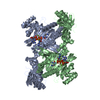


 PDBj
PDBj

 Assembly
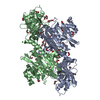
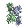
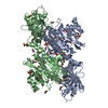
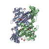
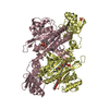
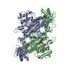
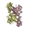
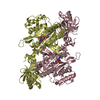
 Chlamydia trachomatis serovar L2b (strain UCH-1/proctitis) (bacteria)


 Type: L-peptide linking / Mass: 147.195 Da / Num. of mol.: 2 / Source method: obtained synthetically / Formula: C6H15N2O2
 Mass: 292.327 Da / Num. of mol.: 2 / Source method: obtained synthetically / Formula: C16H20O5
 Mass: 194.226 Da / Num. of mol.: 2 / Source method: obtained synthetically / Formula: C8H18O5 / Comment: precipitant*YM
 Mass: 62.068 Da / Num. of mol.: 34 / Source method: obtained synthetically / Formula: C2H6O2
 Mass: 106.120 Da / Num. of mol.: 1 / Source method: obtained synthetically / Formula: C4H10O3
 Mass: 22.990 Da / Num. of mol.: 2 / Source method: obtained synthetically / Formula: Na
 Mass: 35.453 Da / Num. of mol.: 1 / Source method: obtained synthetically / Formula: Cl
 Sample preparation
 / Beamline: 21-ID-F / Wavelength: 0.97872 Å
 Processing