[English] 日本語
Yorodumi
- PDB-6nqx: Flagellar protein FcpA from Leptospira biflexa / primitive monocl... -

+
Open data


ID or keywords:

Loading...

-
Basic information

Entry
Database: PDB / ID: 6nqx
TitleFlagellar protein FcpA from Leptospira biflexa / primitive monoclinic form
ComponentsFlagellar coiling protein A
KeywordsPROTEIN FIBRIL / flagellar filament
Function / homologyUncharacterized protein
Function and homology information
Biological speciesLeptospira biflexa serovar Patoc (bacteria)
MethodX-RAY DIFFRACTION / SYNCHROTRON / Resolution: 2.95 Å
AuthorsMechaly, A. / Larrieux, N. / Trajtenberg, F. / Buschiazzo, A.
Citation
Journal: Elife / Year: 2020
Title: An asymmetric sheath controls flagellar supercoiling and motility in the Leptospira spirochete
Authors: Gibson, K. / Trajtenberg, F. / Brady, M. / San Martin, F. / Mechaly, A. / Wunder, E. / Picardeau, M. / Ko, A. / Buschiazzo, A. / Sindelar, C.
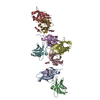
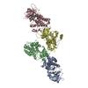
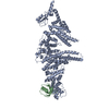
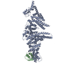
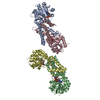
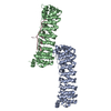
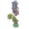
#1: Journal: Acta Crystallogr F Struct Biol Commun / Year: 2017
Title: Crystallization of FcpA from Leptospira, a novel flagellar protein that is essential for pathogenesis.
Authors: Fabiana San Martin / Ariel E Mechaly / Nicole Larrieux / Elsio A Wunder / Albert I Ko / Mathieu Picardeau / Felipe Trajtenberg / Alejandro Buschiazzo /
Abstract: The protein FcpA is a unique component of the flagellar filament of spirochete bacteria belonging to the genus Leptospira. Although it plays an essential role in translational motility and ...The protein FcpA is a unique component of the flagellar filament of spirochete bacteria belonging to the genus Leptospira. Although it plays an essential role in translational motility and pathogenicity, no structures of FcpA homologues are currently available in the PDB. Its three-dimensional structure will unveil the novel motility mechanisms that render pathogenic Leptospira particularly efficient at invading and disseminating within their hosts, causing leptospirosis in humans and animals. FcpA from L. interrogans was purified and crystallized, but despite laborious attempts no useful X ray diffraction data could be obtained. This challenge was solved by expressing a close orthologue from the related saprophytic species L. biflexa. Three different crystal forms were obtained: a primitive and a centred monoclinic form, as well as a hexagonal variant. All forms diffracted X-rays to suitable resolutions for crystallographic analyses, with the hexagonal type typically reaching the highest limits of 2.0 Å and better. A variation of the quick-soaking procedure resulted in an iodide derivative that was instrumental for single-wavelength anomalous diffraction methods.
History
DepositionJan 22, 2019Deposition site: RCSB / Processing site: RCSB
Revision 1.0Jan 29, 2020Provider: repository / Type: Initial release
Revision 1.1Mar 18, 2020Group: Database references / Category: citation
Item: _citation.country / _citation.journal_abbrev ..._citation.country / _citation.journal_abbrev / _citation.journal_id_CSD / _citation.journal_id_ISSN / _citation.journal_volume / _citation.page_first / _citation.title / _citation.year
Revision 1.2Mar 13, 2024Group: Data collection / Database references / Category: chem_comp_atom / chem_comp_bond / database_2
Item: _database_2.pdbx_DOI / _database_2.pdbx_database_accession

-
Structure visualization

Structure viewerMolecule:
MolmilJmol/JSmol

Downloads & links

-
Assembly

Deposited unit
A: Flagellar coiling protein A
B: Flagellar coiling protein A
C: Flagellar coiling protein A
D: Flagellar coiling protein A
hetero molecules


Theoretical massNumber of molelcules
Total (without water)130,09510
Polymers129,5434
Non-polymers5536
Water19811
1


  • Idetical with deposited unit
  • defined by author
  • Evidence: microscopy
TypeNameSymmetry operationNumber
identity operation1_555x,y,z1
Buried area7900 Å2
ΔGint-16 kcal/mol
Surface area50710 Å2
Unit cell
Length a, b, c (Å)85.475, 96.465, 121.138
Angle α, β, γ (deg.)90.000, 105.190, 90.000
Int Tables number4
Space group name H-MP1211

-
Components

#1: Protein
Flagellar coiling protein A


Mass: 32385.668 Da / Num. of mol.: 4 / Fragment: residues 34-300
Source method: isolated from a genetically manipulated source
Source: (gene. exp.) Leptospira biflexa serovar Patoc (strain Patoc 1 / ATCC 23582 / Paris) (bacteria)
Strain: Patoc 1 / ATCC 23582 / Paris / Gene: LEPBI_I0267 / Production host: Escherichia coli (E. coli) / Strain (production host): Rosetta-gami 2 (DE3) / References: UniProt: B0STJ8
#2: Chemical
ChemComp-GOL / GLYCEROL / GLYCERIN / PROPANE-1,2,3-TRIOL / Glycerol


Mass: 92.094 Da / Num. of mol.: 6 / Source method: obtained synthetically / Formula: C3H8O3
#3: Water ChemComp-HOH / water / Water


Mass: 18.015 Da / Num. of mol.: 11 / Source method: isolated from a natural source / Formula: H2O

-
Experimental details

-
Experiment

ExperimentMethod: X-RAY DIFFRACTION / Number of used crystals: 1

-
Sample preparation

CrystalDensity Matthews: 3.72 Å3/Da / Density % sol: 66.94 %
Crystal growTemperature: 293 K / Method: vapor diffusion, hanging drop / Details: PEG 4000, TrisHCl / PH range: 7.5-8.0

-
Data collection

DiffractionMean temperature: 100 K / Serial crystal experiment: N
Diffraction sourceSource: SYNCHROTRON / Site: SOLEIL / Beamline: PROXIMA 1 / Wavelength: 0.97857 Å
DetectorType: DECTRIS PILATUS3 S 6M / Detector: PIXEL / Date: Nov 23, 2013
RadiationProtocol: SINGLE WAVELENGTH / Monochromatic (M) / Laue (L): M / Scattering type: x-ray
Radiation wavelengthWavelength: 0.97857 Å / Relative weight: 1
ReflectionResolution: 2.95→48.23 Å / Num. obs: 39975 / % possible obs: 99.5 % / Redundancy: 3.4 % / Biso Wilson estimate: 102.81 Å2 / CC1/2: 0.999 / Rmerge(I) obs: 0.05 / Rpim(I) all: 0.031 / Rrim(I) all: 0.059 / Net I/σ(I): 17.2
Reflection shell

Diffraction-ID: 1

Resolution (Å)Redundancy (%)Rmerge(I) obsNum. unique obsCC1/2Rpim(I) allRrim(I) all% possible all
2.95-3.073.50.65144850.8420.4050.76899.7
10.64-48.233.20.0168740.9990.0110.0296.8

-
Processing

Software
NameVersionClassification
Aimless0.1.27data scaling
BUSTER2.10.3refinement
PDB_EXTRACT3.24data extraction
TRUNCATEdata reduction
PHASERphasing
RefinementResolution: 2.95→42.7 Å / Cor.coef. Fo:Fc: 0.942 / Cor.coef. Fo:Fc free: 0.925 / SU R Cruickshank DPI: 0.588 / Cross valid method: THROUGHOUT / σ(F): 0 / SU R Blow DPI: 0.514 / SU Rfree Blow DPI: 0.281 / SU Rfree Cruickshank DPI: 0.294
RfactorNum. reflection% reflectionSelection details
Rfree0.222 2005 5.02 %RANDOM
Rwork0.197 ---
obs0.198 39955 99.4 %-
Displacement parametersBiso max: 257.97 Å2 / Biso mean: 115.24 Å2 / Biso min: 42.07 Å2
Baniso -1Baniso -2Baniso -3
1-10.9357 Å20 Å22.5751 Å2
2---23.243 Å20 Å2
3---12.3072 Å2
Refine analyzeLuzzati coordinate error obs: 0.41 Å
Refinement stepCycle: final / Resolution: 2.95→42.7 Å
ProteinNucleic acidLigandSolventTotal
Num. atoms7924 0 36 11 7971
Biso mean--95.98 67.89 -
Num. residues----945
Refine LS restraints
Refine-IDTypeNumberRestraint functionWeightDev ideal
X-RAY DIFFRACTIONt_dihedral_angle_d2995SINUSOIDAL2
X-RAY DIFFRACTIONt_trig_c_planes
X-RAY DIFFRACTIONt_gen_planes1394HARMONIC5
X-RAY DIFFRACTIONt_it8195HARMONIC20
X-RAY DIFFRACTIONt_nbd
X-RAY DIFFRACTIONt_improper_torsion
X-RAY DIFFRACTIONt_pseud_angle
X-RAY DIFFRACTIONt_chiral_improper_torsion1005SEMIHARMONIC5
X-RAY DIFFRACTIONt_sum_occupancies
X-RAY DIFFRACTIONt_utility_distance
X-RAY DIFFRACTIONt_utility_angle
X-RAY DIFFRACTIONt_utility_torsion
X-RAY DIFFRACTIONt_ideal_dist_contact9180SEMIHARMONIC4
X-RAY DIFFRACTIONt_bond_d8195HARMONIC20.01
X-RAY DIFFRACTIONt_angle_deg11037HARMONIC21
X-RAY DIFFRACTIONt_omega_torsion2.4
X-RAY DIFFRACTIONt_other_torsion19.26
LS refinement shellResolution: 2.95→2.97 Å / Rfactor Rfree error: 0 / Total num. of bins used: 50
RfactorNum. reflection% reflection
Rfree0.3235 42 5.25 %
Rwork0.2802 758 -
all0.2828 800 -
obs--99.87 %
Refinement TLS params.

Method: refined / Refine-ID: X-RAY DIFFRACTION

IDL112)L122)L132)L222)L232)L332)S11 (Å °)S12 (Å °)S13 (Å °)S21 (Å °)S22 (Å °)S23 (Å °)S31 (Å °)S32 (Å °)S33 (Å °)T112)T122)T132)T222)T232)T332)Origin x (Å)Origin y (Å)Origin z (Å)
13.00860.20991.64160.53770.80323.23750.0845-0.78460.3017-0.0974-0.09360.0709-0.0856-0.52320.0091-0.337-0.07810.06590.054-0.0153-0.0887-23.7189106.63863.4262
22.20010.64480.37181.74270.88282.1783-0.07520.52930.2728-0.4574-0.036-0.0004-0.3831-0.06970.1111-0.23360.00880.03960.20170.0716-0.2749-35.771876.193128.5842
32.89450.5547-0.09740.92070.05772.06570.053-0.208-0.00020.0643-0.18520.06350.1316-0.48550.1322-0.3014-0.10010.08050.1368-0.0992-0.2048-55.759760.487446.9995
42.53410.70950.90370.87912.27214.35450.56220.2211-0.70880.2213-0.42320.03021.3068-0.7871-0.1390.2792-0.23890.0271-0.1518-0.2801-0.3878-70.058228.273614.2234
Refinement TLS group
IDRefine-IDRefine TLS-IDSelection detailsAuth asym-IDAuth seq-ID
1X-RAY DIFFRACTION1{ A|* }A37 - 292
2X-RAY DIFFRACTION2{ B|* }B63 - 291
3X-RAY DIFFRACTION3{ C|* }C37 - 292
4X-RAY DIFFRACTION4{ D|* }D67 - 291

+
About Yorodumi

-
News

-
Feb 9, 2022. New format data for meta-information of EMDB entries

New format data for meta-information of EMDB entries

  • Version 3 of the EMDB header file is now the official format.
  • The previous official version 1.9 will be removed from the archive.

Related info.:EMDB header

External links:wwPDB to switch to version 3 of the EMDB data model

-
Aug 12, 2020. Covid-19 info

Covid-19 info

URL: https://pdbj.org/emnavi/covid19.php

New page: Covid-19 featured information page in EM Navigator.

Related info.:Covid-19 info / Mar 5, 2020. Novel coronavirus structure data

+
Mar 5, 2020. Novel coronavirus structure data

Novel coronavirus structure data

Related info.:Yorodumi Speices / Aug 12, 2020. Covid-19 info

External links:COVID-19 featured content - PDBj / Molecule of the Month (242):Coronavirus Proteases

+
Jan 31, 2019. EMDB accession codes are about to change! (news from PDBe EMDB page)

EMDB accession codes are about to change! (news from PDBe EMDB page)

  • The allocation of 4 digits for EMDB accession codes will soon come to an end. Whilst these codes will remain in use, new EMDB accession codes will include an additional digit and will expand incrementally as the available range of codes is exhausted. The current 4-digit format prefixed with “EMD-” (i.e. EMD-XXXX) will advance to a 5-digit format (i.e. EMD-XXXXX), and so on. It is currently estimated that the 4-digit codes will be depleted around Spring 2019, at which point the 5-digit format will come into force.
  • The EM Navigator/Yorodumi systems omit the EMD- prefix.

Related info.:Q: What is EMD? / ID/Accession-code notation in Yorodumi/EM Navigator

External links:EMDB Accession Codes are Changing Soon! / Contact to PDBj

+
Jul 12, 2017. Major update of PDB

Major update of PDB

  • wwPDB released updated PDB data conforming to the new PDBx/mmCIF dictionary.
  • This is a major update changing the version number from 4 to 5, and with Remediation, in which all the entries are updated.
  • In this update, many items about electron microscopy experimental information are reorganized (e.g. em_software).
  • Now, EM Navigator and Yorodumi are based on the updated data.

External links:wwPDB Remediation / Enriched Model Files Conforming to OneDep Data Standards Now Available in the PDB FTP Archive

-
Yorodumi

Thousand views of thousand structures

  • Yorodumi is a browser for structure data from EMDB, PDB, SASBDB, etc.
  • This page is also the successor to EM Navigator detail page, and also detail information page/front-end page for Omokage search.
  • The word "yorodu" (or yorozu) is an old Japanese word meaning "ten thousand". "mi" (miru) is to see.

Related info.:EMDB / PDB / SASBDB / Comparison of 3 databanks / Yorodumi Search / Aug 31, 2016. New EM Navigator & Yorodumi / Yorodumi Papers / Jmol/JSmol / Function and homology information / Changes in new EM Navigator and Yorodumi

Read more