 Movie
Movie Controller


+ Open data

- Basic information
| Entry | Database: PDB / ID: 6non | ||||||
|---|---|---|---|---|---|---|---|
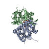
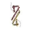
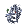
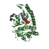
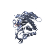
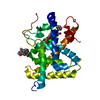
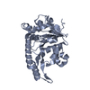
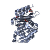
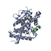
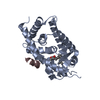
| Title | Structure of Cyanthece apo McdA | ||||||
 Components Components | Cobyrinic acid ac-diamide synthase | ||||||
 Keywords Keywords | DNA BINDING PROTEIN / nonspecific DNA binding protein / Walker box / ParA-like / Carboxysome / McdA / McdB | ||||||
| Function / homology |  Function and homology information Function and homology informationcarbon fixation / nucleoid / Hydrolases; Acting on acid anhydrides; Acting on acid anhydrides to facilitate cellular and subcellular movement / hydrolase activity / DNA binding / ATP binding / cytoplasm Similarity search - Function | ||||||
| Biological species |  Cyanothece (bacteria) Cyanothece (bacteria) | ||||||
| Method |  X-RAY DIFFRACTION / SYNCHROTRON / MAD / Resolution: 2.68 Å X-RAY DIFFRACTION / SYNCHROTRON / MAD / Resolution: 2.68 Å | ||||||
 Authors Authors | Schumacher, M.A. | ||||||
 Citation Citation | Journal: Nucleic Acids Res. / Year: 2019 Title: Structures of maintenance of carboxysome distribution Walker-box McdA and McdB adaptor homologs. Authors: Schumacher, M.A. / Henderson, M. / Zhang, H. | ||||||
| History |
|
- Structure visualization
| Structure viewer | Molecule: MolmilJmol/JSmol |
|---|
- Downloads & links
-Download
| PDBx/mmCIF format | 6non.cif.gz | 208 KB | Display | PDBx/mmCIF format |
|---|---|---|---|---|
| PDB format | pdb6non.ent.gz | 166 KB | Display | PDB format |
| PDBx/mmJSON format | 6non.json.gz | Tree view | PDBx/mmJSON format | |
| Others |  Other downloads Other downloads |
-Validation report
| Arichive directory | https://data.pdbj.org/pub/pdb/validation_reports/no/6nonftp://data.pdbj.org/pub/pdb/validation_reports/no/6non | HTTPS FTP |
|---|
-Related structure data


-Links
 PDBj
PDBj- Assembly
| Deposited unit | 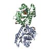
| ||||||||
|---|---|---|---|---|---|---|---|---|---|
| 1 | 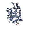
| ||||||||
| 2 | 
| ||||||||
| Unit cell |
|
-Components
| #1: Protein | Mass: 29325.689 Da / Num. of mol.: 2 Source method: isolated from a genetically manipulated source Source: (gene. exp.) Cyanothece (bacteria) / Gene: PCC7424_5529 / Production host: #2: Chemical | ChemComp-ADP / |   Mass: 427.201 Da / Num. of mol.: 1 / Source method: obtained synthetically / Formula: C10H15N5O10P2 / Comment: ADP, energy-carrying molecule*YM Mass: 427.201 Da / Num. of mol.: 1 / Source method: obtained synthetically / Formula: C10H15N5O10P2 / Comment: ADP, energy-carrying molecule*YM#3: Chemical | ChemComp-MG / |   Mass: 24.305 Da / Num. of mol.: 1 / Source method: obtained synthetically / Formula: Mg Mass: 24.305 Da / Num. of mol.: 1 / Source method: obtained synthetically / Formula: Mg#4: Water | ChemComp-HOH / |  Mass: 18.015 Da / Num. of mol.: 79 / Source method: isolated from a natural source / Formula: H2O Mass: 18.015 Da / Num. of mol.: 79 / Source method: isolated from a natural source / Formula: H2OHas protein modification | Y | |
|---|
-Experimental details
-Experiment
| Experiment | Method: X-RAY DIFFRACTION / Number of used crystals: 1 |
|---|
- Sample preparation
| Crystal | Density Matthews: 5.39 Å3/Da / Density % sol: 77.17 % |
|---|---|
| Crystal grow | Temperature: 298 K / Method: vapor diffusion, hanging drop / Details: ammonium sulfate, lithium sulfate |
-Data collection
| Diffraction | Mean temperature: 100 K / Serial crystal experiment: N |
|---|---|
| Diffraction source | Source: SYNCHROTRON / Site: ALS  / Beamline: 8.3.1 / Wavelength: 0.979 Å / Beamline: 8.3.1 / Wavelength: 0.979 Å |
| Detector | Type: DECTRIS PILATUS3 S 6M / Detector: PIXEL / Date: Apr 2, 2018 |
| Radiation | Protocol: MAD / Monochromatic (M) / Laue (L): M / Scattering type: x-ray |
| Radiation wavelength | Wavelength: 0.979 Å / Relative weight: 1 |
| Reflection | Resolution: 2.68→134.5 Å / Num. obs: 61015 / % possible obs: 80.8 % / Observed criterion σ(F): 0 / Observed criterion σ(I): 0 / Redundancy: 4.7 % / Biso Wilson estimate: 57.51 Å2 / CC1/2: 0.997 / Rpim(I) all: 0.045 / Rsym value: 0.07 / Net I/σ(I): 17.6 |
| Reflection shell | Resolution: 2.68→275 Å / Redundancy: 3.5 % / Mean I/σ(I) obs: 2.1 / CC1/2: 0.616 / Rpim(I) all: 0.47 / Rsym value: 0.567 |
-Phasing
| Phasing | Method: MAD |
|---|
- Processing
| Software |
| |||||||||||||||||||||||||||||||||||||||||||||||||||||||||||||||||||||||||||||
|---|---|---|---|---|---|---|---|---|---|---|---|---|---|---|---|---|---|---|---|---|---|---|---|---|---|---|---|---|---|---|---|---|---|---|---|---|---|---|---|---|---|---|---|---|---|---|---|---|---|---|---|---|---|---|---|---|---|---|---|---|---|---|---|---|---|---|---|---|---|---|---|---|---|---|---|---|---|---|
| Refinement | Method to determine structure: MAD / Resolution: 2.68→77.676 Å / SU ML: 0.33 / Cross valid method: THROUGHOUT / σ(F): 1.02 / Phase error: 23.2
| |||||||||||||||||||||||||||||||||||||||||||||||||||||||||||||||||||||||||||||
| Solvent computation | Shrinkage radii: 0.83 Å / VDW probe radii: 1.1 Å / Bsol: 30.912 Å2 / ksol: 0.318 e/Å3 | |||||||||||||||||||||||||||||||||||||||||||||||||||||||||||||||||||||||||||||
| Displacement parameters |
| |||||||||||||||||||||||||||||||||||||||||||||||||||||||||||||||||||||||||||||
| Refinement step | Cycle: LAST / Resolution: 2.68→77.676 Å
| |||||||||||||||||||||||||||||||||||||||||||||||||||||||||||||||||||||||||||||
| Refine LS restraints |
| |||||||||||||||||||||||||||||||||||||||||||||||||||||||||||||||||||||||||||||
| LS refinement shell |
|
