[English] 日本語
Yorodumi
- PDB-6nmj: Crystal Structure of Rat Ric-8A G alpha binding domain, "Paratone... -

+
Open data


ID or keywords:

Loading...

-
Basic information

Entry
Database: PDB / ID: 6nmj
TitleCrystal Structure of Rat Ric-8A G alpha binding domain, "Paratone-N Immersed"
ComponentsResistance to inhibitors of cholinesterase 8 homolog A (C. elegans)
KeywordsSIGNALING PROTEIN / guanine nucleotide exchange factor heterotrimeric G protein alpha subunit G alpha i(1)
Function / homology
Function and homology information


cell-cell adhesion involved in gastrulation / cell migration involved in gastrulation / basement membrane organization / vasculature development / G-protein alpha-subunit binding / adenylate cyclase-inhibiting G protein-coupled receptor signaling pathway / guanyl-nucleotide exchange factor activity / visual learning / in utero embryonic development / plasma membrane / cytoplasm
Similarity search - Function
Synembryn / Guanine nucleotide exchange factor, Ric8 / Guanine nucleotide exchange factor synembryn / Armadillo-like helical / Armadillo-type fold
Similarity search - Domain/homology
Biological speciesRattus norvegicus (Norway rat)
MethodX-RAY DIFFRACTION / SYNCHROTRON / MOLECULAR REPLACEMENT / Resolution: 2.3 Å
AuthorsZeng, B. / Mou, T.C. / Sprang, S.R.
Funding support United States, 2items
OrganizationGrant numberCountry
National Institutes of Health/National Institute of General Medical Sciences (NIH/NIGMS)5R01GM105993 United States
National Institutes of Health/National Institute of General Medical Sciences (NIH/NIGMS)P20GM103546 United States
Citation
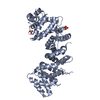
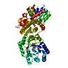
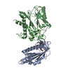
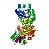
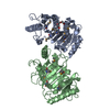
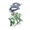
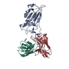
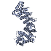
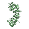
Journal: Structure / Year: 2019
Title: Structure, Function, and Dynamics of the Gα Binding Domain of Ric-8A.
Authors: Baisen Zeng / Tung-Chung Mou / Tzanko I Doukov / Andrea Steiner / Wenxi Yu / Makaia Papasergi-Scott / Gregory G Tall / Franz Hagn / Stephen R Sprang /
Abstract: Ric-8A is a 530-amino acid cytoplasmic molecular chaperone and guanine nucleotide exchange factor (GEF) for i, q, and 12/13 classes of heterortrimeric G protein alpha subunits (Gα). We report the 2. ...Ric-8A is a 530-amino acid cytoplasmic molecular chaperone and guanine nucleotide exchange factor (GEF) for i, q, and 12/13 classes of heterortrimeric G protein alpha subunits (Gα). We report the 2.2-Å crystal structure of the Ric-8A Gα-binding domain with GEF activity, residues 1-452, and is phosphorylated at Ser435 and Thr440. Residues 1-429 adopt a superhelical fold comprised of Armadillo (ARM) and HEAT repeats, and the C terminus is disordered. One of the phosphorylated residues potentially binds to a basic cluster in an ARM motif. Amino acid sequence conservation and published hydrogen-deuterium exchange data indicate repeats 3 through 6 to be a putative Gα-binding surface. Normal mode modeling of small-angle X-ray scattering data indicates that phosphorylation induces relative rotation between repeats 1-4, 5-6, and 7-9. 2D H-N-TROSY spectra of [H,N]-labeled Gαi1 in the presence of R452 reveals chemical shift perturbations of the C terminus and Gαi1 residues involved in nucleotide binding.
History
DepositionJan 11, 2019Deposition site: RCSB / Processing site: RCSB
Revision 1.0Jun 26, 2019Provider: repository / Type: Initial release
Revision 1.1Sep 25, 2019Group: Data collection / Database references / Category: citation / citation_author
Item: _citation.country / _citation.journal_abbrev ..._citation.country / _citation.journal_abbrev / _citation.journal_id_ASTM / _citation.journal_id_CSD / _citation.journal_id_ISSN / _citation.journal_volume / _citation.page_first / _citation.page_last / _citation.pdbx_database_id_DOI / _citation.pdbx_database_id_PubMed / _citation.title / _citation.year
Revision 1.2Jan 1, 2020Group: Author supporting evidence / Category: pdbx_audit_support / Item: _pdbx_audit_support.funding_organization
Revision 1.3Oct 11, 2023Group: Data collection / Database references / Refinement description
Category: chem_comp_atom / chem_comp_bond ...chem_comp_atom / chem_comp_bond / database_2 / pdbx_initial_refinement_model
Item: _database_2.pdbx_DOI / _database_2.pdbx_database_accession

-
Structure visualization

Structure viewerMolecule:
MolmilJmol/JSmol

Downloads & links

-
Assembly

Deposited unit
A: Resistance to inhibitors of cholinesterase 8 homolog A (C. elegans)
B: Resistance to inhibitors of cholinesterase 8 homolog A (C. elegans)


Theoretical massNumber of molelcules
Total (without water)102,3202
Polymers102,3202
Non-polymers00
Water1,72996
1
A: Resistance to inhibitors of cholinesterase 8 homolog A (C. elegans)


Theoretical massNumber of molelcules
Total (without water)51,1601
Polymers51,1601
Non-polymers00
Water181
TypeNameSymmetry operationNumber
identity operation1_555x,y,z1
2
B: Resistance to inhibitors of cholinesterase 8 homolog A (C. elegans)


Theoretical massNumber of molelcules
Total (without water)51,1601
Polymers51,1601
Non-polymers00
Water181
TypeNameSymmetry operationNumber
identity operation1_555x,y,z1
Unit cell
Length a, b, c (Å)63.298, 100.114, 129.959
Angle α, β, γ (deg.)90.00, 90.00, 90.00
Int Tables number19
Space group name H-MP212121

-
Components

#1: Protein Resistance to inhibitors of cholinesterase 8 homolog A (C. elegans) / Ric8a protein / Synembryn-A


Mass: 51159.820 Da / Num. of mol.: 2 / Mutation: F232Y
Source method: isolated from a genetically manipulated source
Source: (gene. exp.) Rattus norvegicus (Norway rat) / Gene: Ric8a, rCG_48458 / Production host: Escherichia coli (E. coli) / References: UniProt: B1H241
#2: Water ChemComp-HOH / water


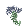
Mass: 18.015 Da / Num. of mol.: 96 / Source method: isolated from a natural source / Formula: H2O

-
Experimental details

-
Experiment

ExperimentMethod: X-RAY DIFFRACTION / Number of used crystals: 1

-
Sample preparation

CrystalDensity Matthews: 2.01 Å3/Da / Density % sol: 38.87 % / Description: Rod
Crystal growTemperature: 293 K / Method: vapor diffusion, sitting drop / Details: 25-30% PEG3350, 0.2 M lithium sulfate, 0.1 M Tris / PH range: 6-9

-
Data collection

DiffractionMean temperature: 100 K / Serial crystal experiment: N
Diffraction sourceSource: SYNCHROTRON / Site: SSRL / Beamline: BL9-2 / Wavelength: 0.98 Å
DetectorType: DECTRIS PILATUS 6M / Detector: PIXEL / Date: Nov 10, 2017
RadiationMonochromator: double crystal Si(111) / Protocol: SINGLE WAVELENGTH / Monochromatic (M) / Laue (L): M / Scattering type: x-ray
Radiation wavelengthWavelength: 0.98 Å / Relative weight: 1
ReflectionResolution: 2.3→39.65 Å / Num. obs: 37176 / % possible obs: 99.11 % / Redundancy: 26.3 % / Biso Wilson estimate: 41 Å2 / CC1/2: 1 / Rpim(I) all: 0.02 / Rrim(I) all: 0.09 / Net I/σ(I): 30.72
Reflection shellResolution: 2.3→2.382 Å / Redundancy: 26.9 % / Mean I/σ(I) obs: 4.55 / Num. unique obs: 3675 / CC1/2: 0.95 / Rpim(I) all: 0.2 / Rrim(I) all: 1.04 / % possible all: 99.19

-
Processing

Software
NameVersionClassification
PHENIX(1.12_2829: ???)refinement
XDSdata reduction
XDSdata scaling
PHASERphasing
RefinementMethod to determine structure: MOLECULAR REPLACEMENT
Starting model: PDB entry 6NMG
Resolution: 2.3→39.655 Å / SU ML: 0.26 / Cross valid method: FREE R-VALUE / σ(F): 1.33 / Phase error: 26.31 / Stereochemistry target values: ML
RfactorNum. reflection% reflection
Rfree0.2593 1171 3.15 %
Rwork0.2077 --
obs0.2094 37129 99.11 %
Solvent computationShrinkage radii: 0.9 Å / VDW probe radii: 1.11 Å / Solvent model: FLAT BULK SOLVENT MODEL
Refinement stepCycle: LAST / Resolution: 2.3→39.655 Å
ProteinNucleic acidLigandSolventTotal
Num. atoms6445 0 0 96 6541
Refine LS restraints
Refine-IDTypeDev idealNumber
X-RAY DIFFRACTIONf_bond_d0.0036546
X-RAY DIFFRACTIONf_angle_d0.578849
X-RAY DIFFRACTIONf_dihedral_angle_d11.2754041
X-RAY DIFFRACTIONf_chiral_restr0.0361051
X-RAY DIFFRACTIONf_plane_restr0.0041134
LS refinement shell
Resolution (Å)Rfactor RfreeNum. reflection RfreeRfactor RworkNum. reflection RworkRefine-ID% reflection obs (%)
2.3-2.40470.33591440.24674419X-RAY DIFFRACTION99
2.4047-2.53150.30871430.24334403X-RAY DIFFRACTION98
2.5315-2.690.24871450.23074462X-RAY DIFFRACTION100
2.69-2.89770.27191460.2314464X-RAY DIFFRACTION100
2.8977-3.18920.27551460.23444480X-RAY DIFFRACTION99
3.1892-3.65040.2931450.21264470X-RAY DIFFRACTION99
3.6504-4.5980.21981480.18174555X-RAY DIFFRACTION99
4.598-39.66080.24791540.19454705X-RAY DIFFRACTION99

+
About Yorodumi

-
News

-
Feb 9, 2022. New format data for meta-information of EMDB entries

New format data for meta-information of EMDB entries

  • Version 3 of the EMDB header file is now the official format.
  • The previous official version 1.9 will be removed from the archive.

Related info.:EMDB header

External links:wwPDB to switch to version 3 of the EMDB data model

-
Aug 12, 2020. Covid-19 info

Covid-19 info

URL: https://pdbjlvh1.pdbj.org/emnavi/covid19.php

New page: Covid-19 featured information page in EM Navigator.

Related info.:Covid-19 info / Mar 5, 2020. Novel coronavirus structure data

+
Mar 5, 2020. Novel coronavirus structure data

Novel coronavirus structure data

Related info.:Yorodumi Speices / Aug 12, 2020. Covid-19 info

External links:COVID-19 featured content - PDBj / Molecule of the Month (242):Coronavirus Proteases

+
Jan 31, 2019. EMDB accession codes are about to change! (news from PDBe EMDB page)

EMDB accession codes are about to change! (news from PDBe EMDB page)

  • The allocation of 4 digits for EMDB accession codes will soon come to an end. Whilst these codes will remain in use, new EMDB accession codes will include an additional digit and will expand incrementally as the available range of codes is exhausted. The current 4-digit format prefixed with “EMD-” (i.e. EMD-XXXX) will advance to a 5-digit format (i.e. EMD-XXXXX), and so on. It is currently estimated that the 4-digit codes will be depleted around Spring 2019, at which point the 5-digit format will come into force.
  • The EM Navigator/Yorodumi systems omit the EMD- prefix.

Related info.:Q: What is EMD? / ID/Accession-code notation in Yorodumi/EM Navigator

External links:EMDB Accession Codes are Changing Soon! / Contact to PDBj

+
Jul 12, 2017. Major update of PDB

Major update of PDB

  • wwPDB released updated PDB data conforming to the new PDBx/mmCIF dictionary.
  • This is a major update changing the version number from 4 to 5, and with Remediation, in which all the entries are updated.
  • In this update, many items about electron microscopy experimental information are reorganized (e.g. em_software).
  • Now, EM Navigator and Yorodumi are based on the updated data.

External links:wwPDB Remediation / Enriched Model Files Conforming to OneDep Data Standards Now Available in the PDB FTP Archive

-
Yorodumi

Thousand views of thousand structures

  • Yorodumi is a browser for structure data from EMDB, PDB, SASBDB, etc.
  • This page is also the successor to EM Navigator detail page, and also detail information page/front-end page for Omokage search.
  • The word "yorodu" (or yorozu) is an old Japanese word meaning "ten thousand". "mi" (miru) is to see.

Related info.:EMDB / PDB / SASBDB / Comparison of 3 databanks / Yorodumi Search / Aug 31, 2016. New EM Navigator & Yorodumi / Yorodumi Papers / Jmol/JSmol / Function and homology information / Changes in new EM Navigator and Yorodumi

Read more