 Movie
Movie Controller

[English] 日本語
 Yorodumi
- SASDFB5: Phosphorylated Resistance to inhibitors of cholinesterase 8 homol... -
+ Open data

- Basic information
| Entry |  |
|---|---|
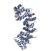
 Sample Sample | Phosphorylated Resistance to inhibitors of cholinesterase 8 homolog A, Ric-8A, amino acids 1-452 (Rattus norvegicus)
|
| Function / homology |  Function and homology information Function and homology informationcell-cell adhesion involved in gastrulation / cell migration involved in gastrulation / basement membrane organization / vasculature development / response to light stimulus / G-protein alpha-subunit binding / gastrulation / protein folding chaperone / guanyl-nucleotide exchange factor activity / GTPase activator activity ...cell-cell adhesion involved in gastrulation / cell migration involved in gastrulation / basement membrane organization / vasculature development / response to light stimulus / G-protein alpha-subunit binding / gastrulation / protein folding chaperone / guanyl-nucleotide exchange factor activity / GTPase activator activity / visual learning / adenylate cyclase-inhibiting G protein-coupled receptor signaling pathway / cell cortex / in utero embryonic development / G protein-coupled receptor signaling pathway / membrane / plasma membrane / cytoplasm Similarity search - Function |
| Biological species |  |
 Citation Citation | Journal: Structure / Year: 2019 Title: Structure, Function, and Dynamics of the Gα Binding Domain of Ric-8A. Authors: Baisen Zeng / Tung-Chung Mou / Tzanko I Doukov / Andrea Steiner / Wenxi Yu / Makaia Papasergi-Scott / Gregory G Tall / Franz Hagn / Stephen R Sprang /   Abstract: Ric-8A is a 530-amino acid cytoplasmic molecular chaperone and guanine nucleotide exchange factor (GEF) for i, q, and 12/13 classes of heterortrimeric G protein alpha subunits (Gα). We report the 2. ...Ric-8A is a 530-amino acid cytoplasmic molecular chaperone and guanine nucleotide exchange factor (GEF) for i, q, and 12/13 classes of heterortrimeric G protein alpha subunits (Gα). We report the 2.2-Å crystal structure of the Ric-8A Gα-binding domain with GEF activity, residues 1-452, and is phosphorylated at Ser435 and Thr440. Residues 1-429 adopt a superhelical fold comprised of Armadillo (ARM) and HEAT repeats, and the C terminus is disordered. One of the phosphorylated residues potentially binds to a basic cluster in an ARM motif. Amino acid sequence conservation and published hydrogen-deuterium exchange data indicate repeats 3 through 6 to be a putative Gα-binding surface. Normal mode modeling of small-angle X-ray scattering data indicates that phosphorylation induces relative rotation between repeats 1-4, 5-6, and 7-9. 2D H-N-TROSY spectra of [H,N]-labeled Gαi1 in the presence of R452 reveals chemical shift perturbations of the C terminus and Gαi1 residues involved in nucleotide binding. |
 Contact author Contact author |
|
- Structure visualization
- Downloads & links
-Data source
| SASBDB page |  SASDFB5 SASDFB5 |
|---|
-Related structure data


-Models
-Sample
| Sample | Name: Phosphorylated Resistance to inhibitors of cholinesterase 8 homolog A, Ric-8A, amino acids 1-452 (Rattus norvegicus) Specimen concentration: 1 mg/ml |
|---|---|
| Buffer | Name: 25mM HEPES, 150mM NaCl / pH: 8 |
| Entity #1606 | Name: Ric-8A / Type: protein Description: Resistance to inhibitors of cholinesterase 8 homolog A Formula weight: 51.09 / Num. of mol.: 1 / Source: Rattus norvegicus / References: UniProt: B1H241 Sequence: GMEPRAVADA LETGEEDAVT EALRSFNREH SQSFTFDDAQ QEDRKRLAKL LVSVLEQGLS PKHRVTWLQT IRILSRDRSC LDSFASRQSL HALACYADIA ISEEPIPQPP DMDVLLESLK CLCNLVLSSP TAQMLAAEAR LVVRLAERVG LYRKRSYPHE VQFFDLRLLF ...Sequence: GMEPRAVADA LETGEEDAVT EALRSFNREH SQSFTFDDAQ QEDRKRLAKL LVSVLEQGLS PKHRVTWLQT IRILSRDRSC LDSFASRQSL HALACYADIA ISEEPIPQPP DMDVLLESLK CLCNLVLSSP TAQMLAAEAR LVVRLAERVG LYRKRSYPHE VQFFDLRLLF LLTALRTDVR QQLFQELHGV RLLTDALELT LGVAPKENPL VILPAQETER AMEILKVLFN ITFDSVKREV DEEDAALYRY LGTLLRHCVM ADAAGDRTEE FHGHTVNLLG NLPLKCLDVL LALELHEGSL EFMGVNMDVI NALLAFLEKR LHQTHRLKEC VAPVLSVLTE CARMHRPARK FLKAQVLPPL RDVRTRPEVG DLLRNKLVRL MTHLDTDVKR VAAEFLFVLC SESVPRFIKY TGYGNAAGLL AARGLMAGGR PEGQYSEDED TDTEEYREAK ASI |
-Experimental information
| Beam | Instrument name: Stanford Synchrotron Radiation Lightsource (SSRL) BL4-2 City: Menlo Park, CA / 国: USA / Type of source: X-ray synchrotron / Wavelength: 0.1127 Å / Dist. spec. to detc.: 1.7 mm | ||||||||||||||||||||||||||||||
|---|---|---|---|---|---|---|---|---|---|---|---|---|---|---|---|---|---|---|---|---|---|---|---|---|---|---|---|---|---|---|---|
| Detector | Name: Rayonix MX225-HE | ||||||||||||||||||||||||||||||
| Scan |  Measurement date: Apr 24, 2018 / Storage temperature: 4 °C / Cell temperature: 23 °C / Exposure time: 1 sec. / Number of frames: 25 / Unit: 1/A /
| ||||||||||||||||||||||||||||||
| Distance distribution function P(R) | 
| ||||||||||||||||||||||||||||||
| Result | 
|
