- PDB-4rho: Crystal structure of a hypothetical protein (BPSL2088) from Burkh... -
+
Open data
ID or keywords:
Loading...
-
Basic information
Entry
Database: PDB / ID: 4rho
Title
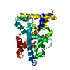
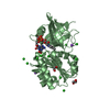
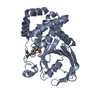
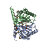
Crystal structure of a hypothetical protein (BPSL2088) from Burkholderia pseudomallei K96243 at 2.25 A resolution
Components
Uncharacterized protein
Keywords
STRUCTURAL GENOMICS / UNKNOWN FUNCTION / New fold / six stranded anit-parallel sheet surrounded by alpha-helices / Imm32 Pfam family / PF15579 / Joint Center for Structural Genomics / JCSG / Protein Structure Initiative / PSI-BIOLOGY
Function / homology
Imm52 family, TsiT-like / Immunity protein 52 / Immunity protein 52 / TRIETHYLENE GLYCOL / Immunity protein 52 domain-containing protein
Function and homology information
Biological species
Burkholderia pseudomallei K96243 (bacteria)
Method
X-RAY DIFFRACTION / SYNCHROTRON / MAD / Resolution: 2.25 Å
Type: MARMOSAIC 325 mm CCD / Detector: CCD / Date: May 3, 2007 Details: Flat mirror (vertical focusing); single crystal Si(111) bent monochromator (ho rizontal focusing)
Radiation
Monochromator: single crystal Si(111) bent / Protocol: MAD / Monochromatic (M) / Laue (L): M / Scattering type: x-ray
Radiation wavelength
ID
Wavelength (Å)
Relative weight
1
0.91837
1
2
0.97937
1
3
0.97895
1
Reflection
Resolution: 2.25→39.071 Å / Num. obs: 24536 / % possible obs: 99 % / Observed criterion σ(I): -3 / Biso Wilson estimate: 33.632 Å2 / Rmerge(I) obs: 0.115 / Net I/σ(I): 6.87
Reflection shell
Resolution (Å)
Rmerge(I) obs
Mean I/σ(I) obs
Num. measured obs
Num. unique obs
Diffraction-ID
% possible all
2.25-2.33
0.549
2.2
7868
2385
1
97.6
2.33-2.42
0.493
2.6
8369
2295
1
99.7
2.42-2.53
0.432
2.8
8923
2455
1
99.6
2.53-2.67
0.319
3.8
9377
2566
1
99.6
2.67-2.83
0.268
4.4
8443
2309
1
99.5
2.83-3.05
0.187
5.6
8859
2446
1
99.2
3.05-3.36
0.132
7.6
8958
2492
1
99.4
3.36-3.84
0.081
10.8
8574
2429
1
99.2
3.84-4.83
0.058
13.6
8755
2494
1
98.6
4.83-39.071
0.053
14.1
9016
2665
1
97.8
-
Phasing
Phasing
Method: MAD
-
Processing
Software
Name
Version
Classification
NB
MolProbity
3beta29
modelbuilding
PDB_EXTRACT
3.1
dataextraction
SHELX
phasing
SHARP
phasing
XSCALE
January10, 2014BUILT=20140307
datascaling
BUSTER-TNT
2.10.0
refinement
XDS
datareduction
SHELXD
phasing
BUSTER
2.10.0
refinement
Refinement
Method to determine structure: MAD / Resolution: 2.25→39.071 Å / Cor.coef. Fo:Fc: 0.9355 / Cor.coef. Fo:Fc free: 0.9254 / Occupancy max: 1 / Occupancy min: 0.33 / Cross valid method: THROUGHOUT / σ(F): 0 Details: 1. A MET-INHIBITION PROTOCOL WAS USED FOR SELENOMETHIONINE INCORPORATION DURING PROTEIN EXPRESSION. THE OCCUPANCY OF THE SE ATOMS IN THE MSE RESIDUES WAS REDUCED TO 0.75 FOR THE REDUCED ...Details: 1. A MET-INHIBITION PROTOCOL WAS USED FOR SELENOMETHIONINE INCORPORATION DURING PROTEIN EXPRESSION. THE OCCUPANCY OF THE SE ATOMS IN THE MSE RESIDUES WAS REDUCED TO 0.75 FOR THE REDUCED SCATTERING POWER DUE TO PARTIAL S-MET INCORPORATION. 2. ATOM RECORD CONTAINS SUM OF TLS AND RESIDUAL B FACTORS. ANISOU RECORD CONTAINS SUM OF TLS AND RESIDUAL U FACTORS. 3. THE MAD PHASES WERE USED AS RESTRAINTS DURING REFINEMENT. 4. NCS RESTRAINTS WERE APPLIED USING BUSTER'S LSSR RESTRAINT REPRESENTATION (-AUTONCS). 5.A POLYETHYLENE GLYCOL FRAGMENTS (PGE) WERE MODELED INTO THE STRUCTURE.
In the structure databanks used in Yorodumi, some data are registered as the other names, "COVID-19 virus" and "2019-nCoV". Here are the details of the virus and the list of structure data.
Jan 31, 2019. EMDB accession codes are about to change! (news from PDBe EMDB page)
EMDB accession codes are about to change! (news from PDBe EMDB page)
The allocation of 4 digits for EMDB accession codes will soon come to an end. Whilst these codes will remain in use, new EMDB accession codes will include an additional digit and will expand incrementally as the available range of codes is exhausted. The current 4-digit format prefixed with “EMD-” (i.e. EMD-XXXX) will advance to a 5-digit format (i.e. EMD-XXXXX), and so on. It is currently estimated that the 4-digit codes will be depleted around Spring 2019, at which point the 5-digit format will come into force.
The EM Navigator/Yorodumi systems omit the EMD- prefix.
Related info.:Q: What is EMD? / ID/Accession-code notation in Yorodumi/EM Navigator
Yorodumi is a browser for structure data from EMDB, PDB, SASBDB, etc.
This page is also the successor to EM Navigator detail page, and also detail information page/front-end page for Omokage search.
The word "yorodu" (or yorozu) is an old Japanese word meaning "ten thousand". "mi" (miru) is to see.
Related info.:EMDB / PDB / SASBDB / Comparison of 3 databanks / Yorodumi Search / Aug 31, 2016. New EM Navigator & Yorodumi / Yorodumi Papers / Jmol/JSmol / Function and homology information / Changes in new EM Navigator and Yorodumi
 Movie
Movie Controller

 Yorodumi
 Open data
 Basic information
 Components
 Keywords
 Function and homology information
 Burkholderia pseudomallei K96243 (bacteria)
 X-RAY DIFFRACTION /
  Authors
 Citation
 Structure visualization
 Downloads & links
 Other downloads
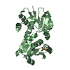


 PDBj
PDBj Assembly
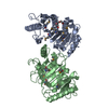


 Mass: 150.173 Da / Num. of mol.: 2 / Source method: obtained synthetically / Formula: C6H14O4
 Mass: 18.015 Da / Num. of mol.: 104 / Source method: isolated from a natural source / Formula: H2O
 Sample preparation
 / Beamline: BL11-1 / Wavelength: 0.91837,0.97937,0.97895
 Processing