| Entry | Database: PDB / ID: 6nji
|
|---|
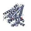
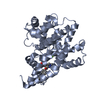
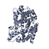
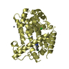
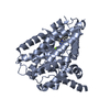
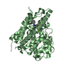
| Title | Crystal Structure of the PDE4D Catalytic Domain and UCR2 Regulatory Helix with T-49 |
|---|
 Components Components | cAMP-specific 3',5'-cyclic phosphodiesterase 4D |
|---|
 Keywords Keywords | HYDROLASE/HYDROLASE INHIBITOR / PDE4D / CAMP-SPECIFIC 3'5'-CYCLIC PHOSPHODIESTERASE 4D / UCR2 / cAMP / HYDROLASE / HYDROLASE-HYDROLASE INHIBITOR complex |
|---|
| Function / homology |  Function and homology information Function and homology information
signaling receptor regulator activity / negative regulation of relaxation of cardiac muscle / negative regulation of heart contraction / 3',5'-cyclic-AMP phosphodiesterase / positive regulation of interleukin-5 production / negative regulation of adenylate cyclase-activating G protein-coupled receptor signaling pathway / regulation of cardiac muscle cell contraction / establishment of endothelial barrier / regulation of calcium ion transmembrane transport via high voltage-gated calcium channel / heterocyclic compound binding ...signaling receptor regulator activity / negative regulation of relaxation of cardiac muscle / negative regulation of heart contraction / 3',5'-cyclic-AMP phosphodiesterase / positive regulation of interleukin-5 production / negative regulation of adenylate cyclase-activating G protein-coupled receptor signaling pathway / regulation of cardiac muscle cell contraction / establishment of endothelial barrier / regulation of calcium ion transmembrane transport via high voltage-gated calcium channel / heterocyclic compound binding / beta-2 adrenergic receptor binding / voltage-gated calcium channel complex / adrenergic receptor signaling pathway / cAMP catabolic process / 3',5'-cyclic-nucleotide phosphodiesterase activity / 3',5'-cyclic-GMP phosphodiesterase activity / regulation of cell communication by electrical coupling involved in cardiac conduction / 3',5'-cyclic-AMP phosphodiesterase activity / DARPP-32 events / cAMP binding / positive regulation of heart rate / negative regulation of cAMP/PKA signal transduction / regulation of release of sequestered calcium ion into cytosol by sarcoplasmic reticulum / positive regulation of interleukin-2 production / cellular response to epinephrine stimulus / calcium channel complex / regulation of heart rate / Turbulent (oscillatory, disturbed) flow shear stress activates signaling by PIEZO1 and integrins in endothelial cells / cellular response to cAMP / calcium channel regulator activity / positive regulation of type II interferon production / T cell receptor signaling pathway / ATPase binding / nuclear membrane / scaffold protein binding / G alpha (s) signalling events / transmembrane transporter binding / cilium / apical plasma membrane / centrosome / enzyme binding / nucleoplasm / membrane / metal ion binding / plasma membrane / cytosolSimilarity search - Function Phosphodiesterase 4 upstream conserved regions (UCR) / Phosphodiesterase 4 upstream conserved regions (UCR) / Catalytic domain of cyclic nucleotide phosphodiesterase 4b2b / 3'5'-cyclic nucleotide phosphodiesterase, catalytic domain / 3'5'-cyclic nucleotide phosphodiesterase / 3'5'-cyclic nucleotide phosphodiesterase, catalytic domain / 3'5'-cyclic nucleotide phosphodiesterase, conserved site / 3'5'-cyclic nucleotide phosphodiesterase, catalytic domain superfamily / 3'5'-cyclic nucleotide phosphodiesterase / 3'5'-cyclic nucleotide phosphodiesterase domain signature. ...Phosphodiesterase 4 upstream conserved regions (UCR) / Phosphodiesterase 4 upstream conserved regions (UCR) / Catalytic domain of cyclic nucleotide phosphodiesterase 4b2b / 3'5'-cyclic nucleotide phosphodiesterase, catalytic domain / 3'5'-cyclic nucleotide phosphodiesterase / 3'5'-cyclic nucleotide phosphodiesterase, catalytic domain / 3'5'-cyclic nucleotide phosphodiesterase, conserved site / 3'5'-cyclic nucleotide phosphodiesterase, catalytic domain superfamily / 3'5'-cyclic nucleotide phosphodiesterase / 3'5'-cyclic nucleotide phosphodiesterase domain signature. / 3'5'-cyclic nucleotide phosphodiesterase domain profile. / Orthogonal Bundle / Mainly AlphaSimilarity search - Domain/homology |
|---|
| Biological species |  Homo sapiens (human) Homo sapiens (human) |
|---|
| Method |  X-RAY DIFFRACTION / SYNCHROTRON / MOLECULAR REPLACEMENT / Resolution: 2.45 Å X-RAY DIFFRACTION / SYNCHROTRON / MOLECULAR REPLACEMENT / Resolution: 2.45 Å |
|---|
 Authors Authors | Fox III, D. / Fairman, J.W. / Gurney, M.E. |
|---|
| Funding support |  United States, 1items United States, 1items | Organization | Grant number | Country |
|---|
| National Institutes of Health/National Institute of Neurological Disorders and Stroke (NIH/NINDS) | NS078034 | United States |
|
|---|
 Citation Citation | Journal: J.Med.Chem. / Year: 2019
Title: Design and Synthesis of Selective Phosphodiesterase 4D (PDE4D) Allosteric Inhibitors for the Treatment of Fragile X Syndrome and Other Brain Disorders.
Authors: Gurney, M.E. / Nugent, R.A. / Mo, X. / Sindac, J.A. / Hagen, T.J. / Fox III, D. / O'Donnell, J.M. / Zhang, C. / Xu, Y. / Zhang, H.T. / Groppi, V.E. / Bailie, M. / White, R.E. / Romero, D.L. ...Authors: Gurney, M.E. / Nugent, R.A. / Mo, X. / Sindac, J.A. / Hagen, T.J. / Fox III, D. / O'Donnell, J.M. / Zhang, C. / Xu, Y. / Zhang, H.T. / Groppi, V.E. / Bailie, M. / White, R.E. / Romero, D.L. / Vellekoop, A.S. / Walker, J.R. / Surman, M.D. / Zhu, L. / Campbell, R.F. |
|---|
| History | | Deposition | Jan 3, 2019 | Deposition site: RCSB / Processing site: RCSB |
|---|
| Revision 1.0 | May 8, 2019 | Provider: repository / Type: Initial release |
|---|
| Revision 1.1 | Jun 5, 2019 | Group: Data collection / Database references / Category: citation / citation_author
Item: _citation.journal_volume / _citation.page_first ..._citation.journal_volume / _citation.page_first / _citation.page_last / _citation_author.identifier_ORCID |
|---|
| Revision 1.2 | Dec 18, 2019 | Group: Author supporting evidence / Category: pdbx_audit_support / Item: _pdbx_audit_support.funding_organization |
|---|
| Revision 1.3 | Mar 13, 2024 | Group: Data collection / Database references / Derived calculations
Category: chem_comp_atom / chem_comp_bond ...chem_comp_atom / chem_comp_bond / database_2 / pdbx_struct_conn_angle / struct_conn
Item: _database_2.pdbx_DOI / _database_2.pdbx_database_accession ..._database_2.pdbx_DOI / _database_2.pdbx_database_accession / _pdbx_struct_conn_angle.ptnr1_auth_seq_id / _pdbx_struct_conn_angle.ptnr3_auth_seq_id / _pdbx_struct_conn_angle.value / _struct_conn.pdbx_dist_value / _struct_conn.ptnr1_auth_asym_id / _struct_conn.ptnr1_auth_comp_id / _struct_conn.ptnr1_auth_seq_id / _struct_conn.ptnr1_label_asym_id / _struct_conn.ptnr1_label_atom_id / _struct_conn.ptnr1_label_comp_id / _struct_conn.ptnr1_label_seq_id / _struct_conn.ptnr2_auth_asym_id / _struct_conn.ptnr2_auth_comp_id / _struct_conn.ptnr2_auth_seq_id / _struct_conn.ptnr2_label_asym_id / _struct_conn.ptnr2_label_atom_id / _struct_conn.ptnr2_label_comp_id |
|---|
|
|---|
 Movie
Movie Controller

 Yorodumi
 Open data
 Basic information
 Structure visualization
 Downloads & links
 Other downloads


 PDBj
PDBj


 Assembly
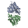
 Trichoplusia ni (cabbage looper)
 Mass: 65.409 Da / Num. of mol.: 2 / Source method: obtained synthetically / Formula: Zn
 Mass: 24.305 Da / Num. of mol.: 2 / Source method: obtained synthetically / Formula: Mg
 Mass: 354.833 Da / Num. of mol.: 2 / Source method: obtained synthetically / Formula: C19H19ClN4O
 Mass: 18.015 Da / Num. of mol.: 69 / Source method: isolated from a natural source / Formula: H2O
 Sample preparation
 Processing