Movie
Movie Controller

[English] 日本語
 Yorodumi
- PDB-6neq: Structure of human mitochondrial translation initiation factor 3 ... -
+ Open data

- Basic information
| Entry | Database: PDB / ID: 6neq | |||||||||
|---|---|---|---|---|---|---|---|---|---|---|
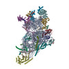
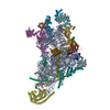
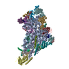
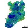
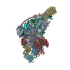
| Title | Structure of human mitochondrial translation initiation factor 3 bound to the small ribosomal subunit-Class-II | |||||||||
 Components Components |
| |||||||||
 Keywords Keywords | RIBOSOMAL PROTEIN / Human mitochondrial translation Initiation Factor 3 | |||||||||
| Function / homology |  Function and homology information Function and homology informationMitochondrial translation elongation / Mitochondrial ribosome-associated quality control / mitochondrial translational initiation / peptide biosynthetic process / translation factor activity, RNA binding / mitochondrial ribosome assembly / Mitochondrial translation initiation / Mitochondrial translation termination / mitochondrial ribosome / ribosome disassembly ...Mitochondrial translation elongation / Mitochondrial ribosome-associated quality control / mitochondrial translational initiation / peptide biosynthetic process / translation factor activity, RNA binding / mitochondrial ribosome assembly / Mitochondrial translation initiation / Mitochondrial translation termination / mitochondrial ribosome / ribosome disassembly / mitochondrial small ribosomal subunit / mitochondrial translation / Mitochondrial protein degradation / ribosomal small subunit binding / translation initiation factor activity / fibrillar center / kinase activity / regulation of translation / ribosome binding / small ribosomal subunit / small ribosomal subunit rRNA binding / tRNA binding / cell population proliferation / mitochondrial inner membrane / rRNA binding / structural constituent of ribosome / ribosome / translation / ribonucleoprotein complex / protein domain specific binding / mRNA binding / apoptotic process / nucleolus / mitochondrion / RNA binding / nucleoplasm / cytosol Similarity search - Function | |||||||||
| Biological species |   Homo sapiens (human) Homo sapiens (human) | |||||||||
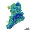
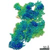
| Method | ELECTRON MICROSCOPY / single particle reconstruction / cryo EM / Resolution: 3.32 Å | |||||||||
 Authors Authors | Sharma, M. / Koripella, R. / Agrawal, R. | |||||||||
| Funding support |  United States, 2items United States, 2items
| |||||||||
 Citation Citation | Journal: iScience / Year: 2019 Title: Structure of Human Mitochondrial Translation Initiation Factor 3 Bound to the Small Ribosomal Subunit. Authors: Ravi K Koripella / Manjuli R Sharma / Md Emdadul Haque / Paul Risteff / Linda L Spremulli / Rajendra K Agrawal / Abstract: The human mitochondrial translational initiation factor 3 (IF3) carries mitochondrial-specific amino acid extensions at both its N and C termini (N- and C-terminal extensions [NTE and CTE, ...The human mitochondrial translational initiation factor 3 (IF3) carries mitochondrial-specific amino acid extensions at both its N and C termini (N- and C-terminal extensions [NTE and CTE, respectively]), when compared with its eubacterial counterpart. Here we present 3.3- to 3.5-Å-resolution cryoelectron microscopic structures of the mammalian 28S mitoribosomal subunit in complex with human IF3. Unique contacts observed between the 28S subunit and N-terminal domain of IF3 explain its unusually high affinity for the 28S subunit, whereas the position of the mito-specific NTE suggests NTE's role in binding of initiator tRNA to the 28S subunit. The location of the C-terminal domain (CTD) clarifies its anti-association activity, whereas the orientation of the mito-specific CTE provides a mechanistic explanation for its role in destabilizing initiator tRNA in the absence of mRNA. Furthermore, our structure hints at a possible role of the CTD in recruiting leaderless mRNAs for translation initiation. Our findings highlight unique features of IF3 in mitochondrial translation initiation. | |||||||||
| History |
|
- Structure visualization
| Movie |
Movie viewer |
|---|---|
| Structure viewer | Molecule: MolmilJmol/JSmol |
 UCSF Chimera
- Downloads & links
-Download
| PDBx/mmCIF format | 6neq.cif.gz | 1.4 MB | Display | PDBx/mmCIF format |
|---|---|---|---|---|
| PDB format | pdb6neq.ent.gz | 1.1 MB | Display | PDB format |
| PDBx/mmJSON format | 6neq.json.gz | Tree view | PDBx/mmJSON format | |
| Others |  Other downloads Other downloads |
-Validation report
| Arichive directory | https://data.pdbj.org/pub/pdb/validation_reports/ne/6neqftp://data.pdbj.org/pub/pdb/validation_reports/ne/6neq | HTTPS FTP |
|---|
-Related structure data
| Related structure data | 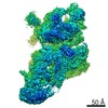 9358MC 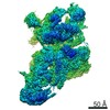 9362C 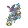 6nf8C M: map data used to model this data C: citing same article ( |
|---|---|
| Similar structure data |

-Links
 PDBj
PDBj


- Assembly
| Deposited unit | 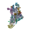
|
|---|---|
| 1 |
|
-Components
-RNA chain , 1 types, 1 molecules A
| #1: RNA chain | Mass: 306932.719 Da / Num. of mol.: 1 / Source method: isolated from a natural source / Source: (natural) |
|---|
+28S ribosomal protein ... , 26 types, 26 molecules CELPQacdejpGIJNhikBFKORUbf
-Protein , 5 types, 5 molecules gmonz
| #17: Protein | Mass: 40505.719 Da / Num. of mol.: 1 / Source method: isolated from a natural source / Source: (natural) |
|---|---|
| #21: Protein | Mass: 13581.974 Da / Num. of mol.: 1 / Source method: isolated from a natural source / Source: (natural) |
| #22: Protein | Mass: 60587.832 Da / Num. of mol.: 1 / Source method: isolated from a natural source / Source: (natural) |
| #31: Protein | Mass: 22968.365 Da / Num. of mol.: 1 / Source method: isolated from a natural source / Source: (natural) |
| #32: Protein | Mass: 28213.160 Da / Num. of mol.: 1 / Fragment: residues 32-278 / Source method: isolated from a natural source / Source: (natural) Homo sapiens (human) / References: UniProt: Q9H2K0 |
-Details
| Has protein modification | Y |
|---|
-Experimental details
-Experiment
| Experiment | Method: ELECTRON MICROSCOPY |
|---|---|
| EM experiment | Aggregation state: PARTICLE / 3D reconstruction method: single particle reconstruction |
- Sample preparation
| Component | Name: Structure of human mitochondrial translation initiation factor 3 bound to the small ribosomal subunit Type: RIBOSOME Details: Structure of human mitochondrial translation initiation factor 3 bound to the small ribosomal subunit Entity ID: all / Source: NATURAL | ||||||||||||
|---|---|---|---|---|---|---|---|---|---|---|---|---|---|
| Source (natural) |
| ||||||||||||
| Buffer solution | pH: 7.6 | ||||||||||||
| Specimen | Embedding applied: NO / Shadowing applied: NO / Staining applied: NO / Vitrification applied: YES | ||||||||||||
| Specimen support | Details: unspecified | ||||||||||||
| Vitrification | Cryogen name: ETHANE |
- Electron microscopy imaging
| Experimental equipment |  Model: Titan Krios / Image courtesy: FEI Company |
|---|---|
| Microscopy | Model: FEI TITAN KRIOS |
| Electron gun | Electron source: OTHER / Accelerating voltage: 300 kV / Illumination mode: OTHER |
| Electron lens | Mode: OTHER |
| Image recording | Electron dose: 70 e/Å2 / Film or detector model: GATAN K2 SUMMIT (4k x 4k) |
- Processing
| EM software |
| ||||||||||||
|---|---|---|---|---|---|---|---|---|---|---|---|---|---|
| CTF correction | Type: NONE | ||||||||||||
| 3D reconstruction | Resolution: 3.32 Å / Resolution method: FSC 0.143 CUT-OFF / Num. of particles: 198355 / Symmetry type: POINT | ||||||||||||
| Atomic model building | B value: 97 / Protocol: OTHER | ||||||||||||
| Atomic model building | PDB-ID: 3JD5 Accession code: 3JD5 / Source name: PDB / Type: experimental model |