 Movie
Movie Controller


+ Open data

- Basic information
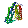
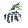
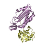
| Entry | Database: PDB / ID: 6nep | ||||||
|---|---|---|---|---|---|---|---|
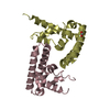
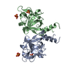
| Title | Structure of human PACRG-MEIG1 complex | ||||||
 Components Components |
| ||||||
 Keywords Keywords | STRUCTURAL PROTEIN / Microtubule / axoneme / primary cilia / flagella | ||||||
| Function / homology |  Function and homology information Function and homology informationaxonemal B tubule inner sheath / axonemal microtubule / manchette / flagellated sperm motility / spermatid development / cellular response to unfolded protein / glial cell projection / alpha-tubulin binding / beta-tubulin binding / heat shock protein binding ...axonemal B tubule inner sheath / axonemal microtubule / manchette / flagellated sperm motility / spermatid development / cellular response to unfolded protein / glial cell projection / alpha-tubulin binding / beta-tubulin binding / heat shock protein binding / Hsp70 protein binding / Hsp90 protein binding / G protein-coupled receptor binding / intracellular protein localization / sperm midpiece / protein-folding chaperone binding / actin binding / cell body / spermatogenesis / vesicle / cell differentiation / neuron projection / ubiquitin protein ligase binding / nucleus / cytosol Similarity search - Function | ||||||
| Biological species |  Homo sapiens (human) Homo sapiens (human) | ||||||
| Method |  X-RAY DIFFRACTION / SYNCHROTRON / SAD / Resolution: 2.09762493085 Å X-RAY DIFFRACTION / SYNCHROTRON / SAD / Resolution: 2.09762493085 Å | ||||||
 Authors Authors | Khan, N. / Croteau, N. / Pelletier, D. / Veyron, S. / Trempe, J.F. | ||||||
| Funding support |  Canada, 1items Canada, 1items
| ||||||
 Citation Citation | Journal: Biorxiv / Year: 2019 Title: Crystal structure of human PACRG in complex with MEIG1 Authors: Khan, N. / Pelletier, D. / Veyron, S. / Croteau, N. / Ichikawa, M. / Black, C. / Khalifa, A.A.Z. / Chaaban, S. / Kurinov, I. / Brouhard, G. / Bui, K.H. / Trempe, J.F. | ||||||
| History |
|
- Structure visualization
| Structure viewer | Molecule: MolmilJmol/JSmol |
|---|
- Downloads & links
-Download
| PDBx/mmCIF format | 6nep.cif.gz | 127.2 KB | Display | PDBx/mmCIF format |
|---|---|---|---|---|
| PDB format | pdb6nep.ent.gz | 93.7 KB | Display | PDB format |
| PDBx/mmJSON format | 6nep.json.gz | Tree view | PDBx/mmJSON format | |
| Others |  Other downloads Other downloads |
-Validation report
| Arichive directory | https://data.pdbj.org/pub/pdb/validation_reports/ne/6nepftp://data.pdbj.org/pub/pdb/validation_reports/ne/6nep | HTTPS FTP |
|---|
-Related structure data


-Links
 PDBj
PDBj
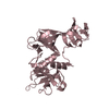
- Assembly
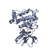
| Deposited unit | 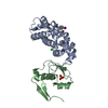
| ||||||||||||
|---|---|---|---|---|---|---|---|---|---|---|---|---|---|
| 1 | 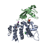
| ||||||||||||
| Unit cell |
|
-Components
| #1: Protein | Mass: 22298.510 Da / Num. of mol.: 1 / Mutation: Deletion 1-69, Y189PHI Source method: isolated from a genetically manipulated source Details: Generated from GST-fusion, and cleaved with 3C protease, leaving a GPLGS linker at N-terminus. Tyrosine 189 was mutated in Iodo-phenylalanine in order to incorporate anomalous scattering atom for phasing. Source: (gene. exp.) Homo sapiens (human) / Gene: PACRG, GLUP / Plasmid: pGEX-6P1 / Production host:  |
|---|---|
| #2: Protein | Mass: 11226.584 Da / Num. of mol.: 1 Source method: isolated from a genetically manipulated source Details: Full-length MEIG1 expressed as His-tagged protein, cleaved with the 3C protease, leaving GPLGS at N-terminus Source: (gene. exp.) Homo sapiens (human) / Gene: MEIG1 / Production host: |
| #3: Chemical | ChemComp-CL /   Mass: 35.453 Da / Num. of mol.: 1 / Source method: obtained synthetically / Formula: Cl Mass: 35.453 Da / Num. of mol.: 1 / Source method: obtained synthetically / Formula: Cl |
| #4: Chemical | ChemComp-PO4 /   Mass: 94.971 Da / Num. of mol.: 1 / Source method: obtained synthetically / Formula: PO4 Mass: 94.971 Da / Num. of mol.: 1 / Source method: obtained synthetically / Formula: PO4 |
| #5: Water | ChemComp-HOH /  Mass: 18.015 Da / Num. of mol.: 74 / Source method: isolated from a natural source / Formula: H2O Mass: 18.015 Da / Num. of mol.: 74 / Source method: isolated from a natural source / Formula: H2O |
| Has protein modification | Y |
-Experimental details
-Experiment
| Experiment | Method: X-RAY DIFFRACTION / Number of used crystals: 1 |
|---|
- Sample preparation
| Crystal | Density Matthews: 2.7 Å3/Da / Density % sol: 54.49 % |
|---|---|
| Crystal grow | Temperature: 283 K / Method: vapor diffusion, sitting drop / Details: 18%PEG8K, 60mM KHPO4, 15% Glycerol |
-Data collection
| Diffraction | Mean temperature: 100 K / Serial crystal experiment: N |
|---|---|
| Diffraction source | Source: SYNCHROTRON / Site: APS  / Beamline: 24-ID-C / Wavelength: 1.476 Å / Beamline: 24-ID-C / Wavelength: 1.476 Å |
| Detector | Type: DECTRIS PILATUS 6M-F / Detector: PIXEL / Date: Nov 28, 2018 |
| Radiation | Protocol: SINGLE WAVELENGTH / Monochromatic (M) / Laue (L): M / Scattering type: x-ray |
| Radiation wavelength | Wavelength: 1.476 Å / Relative weight: 1 |
| Reflection | Resolution: 2.09→47.64 Å / Num. obs: 253322 / % possible obs: 99.8 % / Redundancy: 11.3 % / Biso Wilson estimate: 43.8362539878 Å2 / CC1/2: 0.998 / Rmerge(I) obs: 0.093 / Net I/σ(I): 13.7 |
| Reflection shell | Resolution: 2.09→2.16 Å / Redundancy: 5.6 % / Rmerge(I) obs: 1.1 / Mean I/σ(I) obs: 1.1 / Num. unique obs: 9944 / CC1/2: 0.73 / % possible all: 98.4 |
- Processing
| Software |
| ||||||||||||||||||||||||||||||||||||||||||||||||||||||||||||||||||||||||||||||||||||||||||||||||||||||||||||||||
|---|---|---|---|---|---|---|---|---|---|---|---|---|---|---|---|---|---|---|---|---|---|---|---|---|---|---|---|---|---|---|---|---|---|---|---|---|---|---|---|---|---|---|---|---|---|---|---|---|---|---|---|---|---|---|---|---|---|---|---|---|---|---|---|---|---|---|---|---|---|---|---|---|---|---|---|---|---|---|---|---|---|---|---|---|---|---|---|---|---|---|---|---|---|---|---|---|---|---|---|---|---|---|---|---|---|---|---|---|---|---|---|---|---|
| Refinement | Method to determine structure: SAD / Resolution: 2.09762493085→47.6384909553 Å / SU ML: 0.281412881549 / Cross valid method: THROUGHOUT / σ(F): 0.360717156976 / Phase error: 31.4403924482
| ||||||||||||||||||||||||||||||||||||||||||||||||||||||||||||||||||||||||||||||||||||||||||||||||||||||||||||||||
| Solvent computation | Shrinkage radii: 0.9 Å / VDW probe radii: 1.11 Å | ||||||||||||||||||||||||||||||||||||||||||||||||||||||||||||||||||||||||||||||||||||||||||||||||||||||||||||||||
| Displacement parameters | Biso mean: 51.9849979618 Å2 | ||||||||||||||||||||||||||||||||||||||||||||||||||||||||||||||||||||||||||||||||||||||||||||||||||||||||||||||||
| Refinement step | Cycle: LAST / Resolution: 2.09762493085→47.6384909553 Å
| ||||||||||||||||||||||||||||||||||||||||||||||||||||||||||||||||||||||||||||||||||||||||||||||||||||||||||||||||
| Refine LS restraints |
| ||||||||||||||||||||||||||||||||||||||||||||||||||||||||||||||||||||||||||||||||||||||||||||||||||||||||||||||||
| LS refinement shell |
|
