 Movie
Movie Controller

[English] 日本語
 Yorodumi
- PDB-6nc2: AMC011 v4.2 SOSIP Env trimer in complex with fusion peptide targe... -
+ Open data

- Basic information
| Entry | Database: PDB / ID: 6nc2 | ||||||||||||||||||
|---|---|---|---|---|---|---|---|---|---|---|---|---|---|---|---|---|---|---|---|
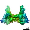
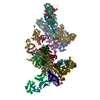
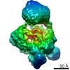
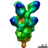
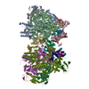
| Title | AMC011 v4.2 SOSIP Env trimer in complex with fusion peptide targeting antibody ACS202 fragment antigen binding | ||||||||||||||||||
 Components Components |
| ||||||||||||||||||
 Keywords Keywords | IMMUNE SYSTEM / HIV-1 Env / SOSIP / trimer / broadly neutralizing antibody / fusion peptide / VIRAL PROTEIN | ||||||||||||||||||
| Function / homology |  Function and homology information Function and homology informationsymbiont-mediated perturbation of host defense response / clathrin-dependent endocytosis of virus by host cell / fusion of virus membrane with host endosome membrane / viral envelope / virion attachment to host cell / host cell plasma membrane / virion membrane / structural molecule activity Similarity search - Function | ||||||||||||||||||
| Biological species |   Human immunodeficiency virus 1 Human immunodeficiency virus 1 Homo sapiens (human) Homo sapiens (human) | ||||||||||||||||||
| Method | ELECTRON MICROSCOPY / single particle reconstruction / cryo EM / Resolution: 5.2 Å | ||||||||||||||||||
 Authors Authors | Cottrell, C.A. / Ozorowski, G. / Yuan, M. / Copps, J. / Wilson, I.A. / Ward, A.B. | ||||||||||||||||||
| Funding support |  United States, 5items United States, 5items
| ||||||||||||||||||
 Citation Citation | Journal: Cell Host Microbe / Year: 2019 Title: Conformational Plasticity in the HIV-1 Fusion Peptide Facilitates Recognition by Broadly Neutralizing Antibodies. Authors: Meng Yuan / Christopher A Cottrell / Gabriel Ozorowski / Marit J van Gils / Sonu Kumar / Nicholas C Wu / Anita Sarkar / Jonathan L Torres / Natalia de Val / Jeffrey Copps / John P Moore / ...Authors: Meng Yuan / Christopher A Cottrell / Gabriel Ozorowski / Marit J van Gils / Sonu Kumar / Nicholas C Wu / Anita Sarkar / Jonathan L Torres / Natalia de Val / Jeffrey Copps / John P Moore / Rogier W Sanders / Andrew B Ward / Ian A Wilson /  Abstract: The fusion peptide (FP) of HIV-1 envelope glycoprotein (Env) is essential for mediating viral entry. Detection of broadly neutralizing antibodies (bnAbs) that interact with the FP has revealed it as ...The fusion peptide (FP) of HIV-1 envelope glycoprotein (Env) is essential for mediating viral entry. Detection of broadly neutralizing antibodies (bnAbs) that interact with the FP has revealed it as a site of vulnerability. We delineate X-ray and cryo-electron microscopy (cryo-EM) structures of bnAb ACS202, from an HIV-infected elite neutralizer, with an FP and with a soluble Env trimer (AMC011 SOSIP.v4.2) derived from the same patient. We show that ACS202 CDRH3 forms a "β strand" interaction with the exposed hydrophobic FP and recognizes a continuous region of gp120, including a conserved N-linked glycan at N88. A cryo-EM structure of another previously identified bnAb VRC34.01 with AMC011 SOSIP.v4.2 shows that it also penetrates through glycans to target the FP. We further demonstrate that the FP can twist and present different conformations for recognition by bnAbs, which enables approach to Env from diverse angles. The variable recognition of FP by bnAbs thus provides insights for vaccine design. | ||||||||||||||||||
| History |
|
- Structure visualization
| Movie |
Movie viewer |
|---|---|
| Structure viewer | Molecule: MolmilJmol/JSmol |
- Downloads & links
-Download
| PDBx/mmCIF format | 6nc2.cif.gz | 1017.6 KB | Display | PDBx/mmCIF format |
|---|---|---|---|---|
| PDB format | pdb6nc2.ent.gz | 820 KB | Display | PDB format |
| PDBx/mmJSON format | 6nc2.json.gz | Tree view | PDBx/mmJSON format | |
| Others |  Other downloads Other downloads |
-Validation report
| Arichive directory | https://data.pdbj.org/pub/pdb/validation_reports/nc/6nc2ftp://data.pdbj.org/pub/pdb/validation_reports/nc/6nc2 | HTTPS FTP |
|---|
-Related structure data
| Related structure data | 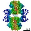 0433MC 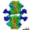 0434C 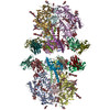 6nc3C 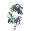 6ncpC M: map data used to model this data C: citing same article ( |
|---|---|
| Similar structure data |

-Links
 PDBj
PDBj


- Assembly
| Deposited unit | 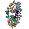
|
|---|---|
| 1 |
|
-Components
-AMC011 v4.2 SOSIP ... , 2 types, 12 molecules ACDEFGBIJKMN
| #1: Protein | Mass: 57668.938 Da / Num. of mol.: 6 / Mutation: H66R, A316W, A501C Source method: isolated from a genetically manipulated source Source: (gene. exp.) Human immunodeficiency virus 1 / Cell line (production host): HEK293F / Production host: Homo sapiens (human)#2: Protein | Mass: 17300.666 Da / Num. of mol.: 6 / Mutation: L543Q, I559P, Q567K, T605C Source method: isolated from a genetically manipulated source Source: (gene. exp.) Human immunodeficiency virus 1 / Cell line (production host): HEK293F / Production host: Homo sapiens (human) / References: UniProt: Q78156*PLUS |
|---|
-Antibody , 2 types, 12 molecules HOPQRSLTUVWX
| #3: Antibody | Mass: 27392.023 Da / Num. of mol.: 6 Source method: isolated from a genetically manipulated source Source: (gene. exp.) Homo sapiens (human) / Cell line (production host): HEK293F / Production host: Homo sapiens (human)#4: Antibody | Mass: 25475.658 Da / Num. of mol.: 6 Source method: isolated from a genetically manipulated source Source: (gene. exp.) Homo sapiens (human) / Cell line (production host): HEK293F / Production host: Homo sapiens (human) |
|---|
-Sugars , 4 types, 54 molecules 
| #5: Polysaccharide | alpha-D-mannopyranose-(1-3)-[alpha-D-mannopyranose-(1-6)]beta-D-mannopyranose-(1-4)-2-acetamido-2- ...alpha-D-mannopyranose-(1-3)-[alpha-D-mannopyranose-(1-6)]beta-D-mannopyranose-(1-4)-2-acetamido-2-deoxy-beta-D-glucopyranose-(1-4)-2-acetamido-2-deoxy-beta-D-glucopyranose Source method: isolated from a genetically manipulated source #6: Polysaccharide | 2-acetamido-2-deoxy-beta-D-glucopyranose-(1-4)-2-acetamido-2-deoxy-beta-D-glucopyranose Source method: isolated from a genetically manipulated source #7: Polysaccharide | alpha-D-mannopyranose-(1-2)-alpha-D-mannopyranose-(1-3)-[alpha-D-mannopyranose-(1-6)]beta-D- ...alpha-D-mannopyranose-(1-2)-alpha-D-mannopyranose-(1-3)-[alpha-D-mannopyranose-(1-6)]beta-D-mannopyranose-(1-4)-2-acetamido-2-deoxy-beta-D-glucopyranose-(1-4)-2-acetamido-2-deoxy-beta-D-glucopyranose Source method: isolated from a genetically manipulated source #8: Sugar | ChemComp-NAG /  Type: D-saccharide, beta linking / Mass: 221.208 Da / Num. of mol.: 30 Type: D-saccharide, beta linking / Mass: 221.208 Da / Num. of mol.: 30Source method: isolated from a genetically manipulated source Formula: C8H15NO6 |
|---|
-Details
| Has protein modification | Y |
|---|
-Experimental details
-Experiment
| Experiment | Method: ELECTRON MICROSCOPY |
|---|---|
| EM experiment | Aggregation state: PARTICLE / 3D reconstruction method: single particle reconstruction |
- Sample preparation
| Component |
| ||||||||||||||||||||||||||||||
|---|---|---|---|---|---|---|---|---|---|---|---|---|---|---|---|---|---|---|---|---|---|---|---|---|---|---|---|---|---|---|---|
| Molecular weight | Value: 0.57 MDa / Experimental value: NO | ||||||||||||||||||||||||||||||
| Source (natural) |
| ||||||||||||||||||||||||||||||
| Source (recombinant) |
| ||||||||||||||||||||||||||||||
| Buffer solution | pH: 7.4 Details: Detergent (DDM) added immediately prior to grid preparation | ||||||||||||||||||||||||||||||
| Buffer component |
| ||||||||||||||||||||||||||||||
| Specimen | Conc.: 5 mg/ml / Embedding applied: NO / Shadowing applied: NO / Staining applied: NO / Vitrification applied: YES Details: SEC purified sample after overnight incubation of molar excess Fab to trimer | ||||||||||||||||||||||||||||||
| Specimen support | Details: Gatan Solarus 950 Plasma system / Grid material: COPPER / Grid mesh size: 400 divisions/in. / Grid type: C-flat-2/2 4C | ||||||||||||||||||||||||||||||
| Vitrification | Instrument: HOMEMADE PLUNGER / Cryogen name: ETHANE |
- Electron microscopy imaging
| Experimental equipment |  Model: Titan Krios / Image courtesy: FEI Company |
|---|---|
| Microscopy | Model: FEI TITAN KRIOS |
| Electron gun | Electron source:  FIELD EMISSION GUN / Accelerating voltage: 300 kV / Illumination mode: FLOOD BEAM FIELD EMISSION GUN / Accelerating voltage: 300 kV / Illumination mode: FLOOD BEAM |
| Electron lens | Mode: BRIGHT FIELD / Nominal magnification: 29000 X / Nominal defocus max: 2500 nm / Nominal defocus min: 1300 nm / Cs: 2.7 mm / C2 aperture diameter: 70 µm / Alignment procedure: COMA FREE |
| Specimen holder | Cryogen: NITROGEN / Specimen holder model: FEI TITAN KRIOS AUTOGRID HOLDER |
| Image recording | Average exposure time: 10 sec. / Electron dose: 92 e/Å2 / Detector mode: COUNTING / Film or detector model: GATAN K2 SUMMIT (4k x 4k) / Num. of real images: 1641 |
| Image scans | Sampling size: 5 µm / Width: 3710 / Height: 3838 / Movie frames/image: 50 |
- Processing
| EM software |
| ||||||||||||||||||||||||||||||||||||
|---|---|---|---|---|---|---|---|---|---|---|---|---|---|---|---|---|---|---|---|---|---|---|---|---|---|---|---|---|---|---|---|---|---|---|---|---|---|
| CTF correction | Type: PHASE FLIPPING AND AMPLITUDE CORRECTION | ||||||||||||||||||||||||||||||||||||
| Symmetry | Point symmetry: D3 (2x3 fold dihedral) | ||||||||||||||||||||||||||||||||||||
| 3D reconstruction | Resolution: 5.2 Å / Resolution method: FSC 0.143 CUT-OFF / Num. of particles: 49878 / Symmetry type: POINT | ||||||||||||||||||||||||||||||||||||
| Atomic model building | Space: REAL |
