[English] 日本語
Yorodumi
- PDB-6nb9: Amyloid-Beta (20-34) with L-isoaspartate 23 -

+
Open data


ID or keywords:

Loading...

-
Basic information

Entry
Database: PDB / ID: 6nb9
TitleAmyloid-Beta (20-34) with L-isoaspartate 23
ComponentsAmyloid-beta A4 protein
KeywordsPROTEIN FIBRIL / protofilament / 2 sub 1 screw symmetry / post-translational modification / isoaspartate
Function / homology
Function and homology information


amyloid-beta complex / negative regulation of presynapse assembly / cytosolic mRNA polyadenylation / collateral sprouting in absence of injury / microglia development / regulation of synapse structure or activity / regulation of Wnt signaling pathway / synaptic assembly at neuromuscular junction / Formyl peptide receptors bind formyl peptides and many other ligands / axo-dendritic transport ...amyloid-beta complex / negative regulation of presynapse assembly / cytosolic mRNA polyadenylation / collateral sprouting in absence of injury / microglia development / regulation of synapse structure or activity / regulation of Wnt signaling pathway / synaptic assembly at neuromuscular junction / Formyl peptide receptors bind formyl peptides and many other ligands / axo-dendritic transport / axon midline choice point recognition / smooth endoplasmic reticulum calcium ion homeostasis / astrocyte activation involved in immune response / NMDA selective glutamate receptor signaling pathway / mating behavior / regulation of spontaneous synaptic transmission / ciliary rootlet / Golgi-associated vesicle / PTB domain binding / Lysosome Vesicle Biogenesis / Insertion of tail-anchored proteins into the endoplasmic reticulum membrane / positive regulation of amyloid fibril formation / neuron remodeling / Deregulated CDK5 triggers multiple neurodegenerative pathways in Alzheimer's disease models / nuclear envelope lumen / COPII-coated ER to Golgi transport vesicle / suckling behavior / signaling receptor activator activity / dendrite development / modulation of excitatory postsynaptic potential / TRAF6 mediated NF-kB activation / presynaptic active zone / positive regulation of protein metabolic process / neuromuscular process controlling balance / Advanced glycosylation endproduct receptor signaling / The NLRP3 inflammasome / negative regulation of long-term synaptic potentiation / regulation of presynapse assembly / regulation of multicellular organism growth / transition metal ion binding / intracellular copper ion homeostasis / negative regulation of neuron differentiation / ECM proteoglycans / spindle midzone / positive regulation of T cell migration / smooth endoplasmic reticulum / Purinergic signaling in leishmaniasis infection / forebrain development / positive regulation of chemokine production / clathrin-coated pit / Notch signaling pathway / protein serine/threonine kinase binding / positive regulation of G2/M transition of mitotic cell cycle / extracellular matrix organization / neuron projection maintenance / Mitochondrial protein degradation / response to interleukin-1 / ionotropic glutamate receptor signaling pathway / positive regulation of mitotic cell cycle / cholesterol metabolic process / axonogenesis / positive regulation of calcium-mediated signaling / dendritic shaft / platelet alpha granule lumen / adult locomotory behavior / positive regulation of glycolytic process / central nervous system development / learning / positive regulation of interleukin-1 beta production / trans-Golgi network membrane / positive regulation of long-term synaptic potentiation / endosome lumen / locomotory behavior / astrocyte activation / Post-translational protein phosphorylation / positive regulation of JNK cascade / microglial cell activation / regulation of long-term neuronal synaptic plasticity / serine-type endopeptidase inhibitor activity / synapse organization / TAK1-dependent IKK and NF-kappa-B activation / positive regulation of non-canonical NF-kappaB signal transduction / neuromuscular junction / visual learning / recycling endosome / positive regulation of interleukin-6 production / Golgi lumen / cognition / neuron cellular homeostasis / positive regulation of inflammatory response / endocytosis / Regulation of Insulin-like Growth Factor (IGF) transport and uptake by Insulin-like Growth Factor Binding Proteins (IGFBPs) / cellular response to amyloid-beta / neuron projection development / G2/M transition of mitotic cell cycle / positive regulation of tumor necrosis factor production / apical part of cell / synaptic vesicle / cell-cell junction / Platelet degranulation
Similarity search - Function
Amyloidogenic glycoprotein, copper-binding / Amyloidogenic glycoprotein, copper-binding domain conserved site / Amyloidogenic glycoprotein, copper-binding domain superfamily / Copper-binding of amyloid precursor, CuBD / Amyloid precursor protein (APP) copper-binding (CuBD) domain signature. / Amyloidogenic glycoprotein, amyloid-beta peptide superfamily / Beta-amyloid peptide (beta-APP) / Amyloidogenic glycoprotein, amyloid-beta peptide / Beta-amyloid precursor protein C-terminal / Amyloidogenic glycoprotein, intracellular domain, conserved site ...Amyloidogenic glycoprotein, copper-binding / Amyloidogenic glycoprotein, copper-binding domain conserved site / Amyloidogenic glycoprotein, copper-binding domain superfamily / Copper-binding of amyloid precursor, CuBD / Amyloid precursor protein (APP) copper-binding (CuBD) domain signature. / Amyloidogenic glycoprotein, amyloid-beta peptide superfamily / Beta-amyloid peptide (beta-APP) / Amyloidogenic glycoprotein, amyloid-beta peptide / Beta-amyloid precursor protein C-terminal / Amyloidogenic glycoprotein, intracellular domain, conserved site / Beta-amyloid precursor protein C-terminus / Amyloid precursor protein (APP) intracellular domain signature. / Amyloidogenic glycoprotein, extracellular / Amyloidogenic glycoprotein, heparin-binding / Amyloidogenic glycoprotein, E2 domain / E2 domain superfamily / Amyloidogenic glycoprotein, heparin-binding domain superfamily / Amyloid A4 N-terminal heparin-binding / E2 domain of amyloid precursor protein / Amyloid precursor protein (APP) E1 domain profile. / Amyloid precursor protein (APP) E2 domain profile. / amyloid A4 / Amyloidogenic glycoprotein / Proteinase inhibitor I2, Kunitz, conserved site / Pancreatic trypsin inhibitor (Kunitz) family signature. / BPTI/Kunitz family of serine protease inhibitors. / Pancreatic trypsin inhibitor Kunitz domain / Kunitz/Bovine pancreatic trypsin inhibitor domain / Pancreatic trypsin inhibitor (Kunitz) family profile. / Pancreatic trypsin inhibitor Kunitz domain superfamily / PH-like domain superfamily
Similarity search - Domain/homology
Amyloid-beta precursor protein
Similarity search - Component
Biological speciesHomo sapiens (human)
MethodELECTRON CRYSTALLOGRAPHY / electron crystallography / AB INITIO PHASING / cryo EM / Resolution: 1.05 Å
Model details
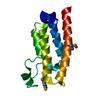
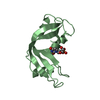
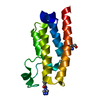
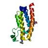
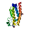
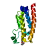
Protofilament structure of Amyloid-beta 20-34 with the age-associated post-translational ...Protofilament structure of Amyloid-beta 20-34 with the age-associated post-translational modification of aspartate isomerization at position 23
AuthorsSawaya, M.R. / Warmack, R.A. / Boyer, D.R. / Zee, C.T. / Richards, L.S. / Cascio, D. / Gonen, T. / Clarke, S.G. / Eisenberg, D.S.
Funding support United States, 4items
OrganizationGrant numberCountry
National Institutes of Health/National Center for Research Resources (NIH/NCRR)5T32GM008496 United States
National Science Foundation (NSF, United States)MCB-1714569 United States
National Institutes of Health/National Center for Research Resources (NIH/NCRR)GM-007185 United States
Howard Hughes Medical Institute (HHMI) United States
CitationJournal: Nat Commun / Year: 2019
Title: Structure of amyloid-β (20-34) with Alzheimer's-associated isomerization at Asp23 reveals a distinct protofilament interface.
Authors: Rebeccah A Warmack / David R Boyer / Chih-Te Zee / Logan S Richards / Michael R Sawaya / Duilio Cascio / Tamir Gonen / David S Eisenberg / Steven G Clarke /
Abstract: Amyloid-β (Aβ) harbors numerous posttranslational modifications (PTMs) that may affect Alzheimer's disease (AD) pathogenesis. Here we present the 1.1 Å resolution MicroED structure of an Aβ 20- ...Amyloid-β (Aβ) harbors numerous posttranslational modifications (PTMs) that may affect Alzheimer's disease (AD) pathogenesis. Here we present the 1.1 Å resolution MicroED structure of an Aβ 20-34 fibril with and without the disease-associated PTM, L-isoaspartate, at position 23 (L-isoAsp23). Both wild-type and L-isoAsp23 protofilaments adopt β-helix-like folds with tightly packed cores, resembling the cores of full-length fibrillar Aβ structures, and both self-associate through two distinct interfaces. One of these is a unique Aβ interface strengthened by the isoaspartyl modification. Powder diffraction patterns suggest a similar structure may be adopted by protofilaments of an analogous segment containing the heritable Iowa mutation, Asp23Asn. Consistent with its early onset phenotype in patients, Asp23Asn accelerates aggregation of Aβ 20-34, as does the L-isoAsp23 modification. These structures suggest that the enhanced amyloidogenicity of the modified Aβ segments may also reduce the concentration required to achieve nucleation and therefore help spur the pathogenesis of AD.
History
DepositionDec 6, 2018Deposition site: RCSB / Processing site: RCSB
Revision 1.0Aug 7, 2019Provider: repository / Type: Initial release
Revision 1.1Nov 20, 2019Group: Author supporting evidence / Category: pdbx_audit_support / Item: _pdbx_audit_support.funding_organization
Revision 1.2Sep 7, 2022Group: Data collection / Database references / Refinement description
Category: database_2 / em_diffraction_shell / refine_hist
Item: _database_2.pdbx_DOI / _database_2.pdbx_database_accession ..._database_2.pdbx_DOI / _database_2.pdbx_database_accession / _em_diffraction_shell.em_diffraction_stats_id / _em_diffraction_shell.id / _refine_hist.pdbx_refine_id
Revision 1.3Nov 13, 2024Group: Data collection / Structure summary
Category: chem_comp_atom / chem_comp_bond ...chem_comp_atom / chem_comp_bond / em_admin / pdbx_entry_details / pdbx_modification_feature
Item: _em_admin.last_update

-
Structure visualization

Movie
  • Biological unit as author_defined_assembly
  • Imaged by Jmol
  • Download
  • Deposited structure unit
  • Imaged by Jmol
  • Download
  • Simplified surface model + fitted atomic model
  • EMDB-0405
  • Imaged by Jmol
  • Download
  • Superimposition on EM map
  • EMDB-0405
  • Imaged by UCSF Chimera
  • Download
Movie viewer
Structure viewerMolecule:
MolmilJmol/JSmol

Downloads & links

-
Assembly

Deposited unit
A: Amyloid-beta A4 protein


Theoretical massNumber of molelcules
Total (without water)1,4921
Polymers1,4921
Non-polymers00
Water724
1
A: Amyloid-beta A4 protein
x 10


Theoretical massNumber of molelcules
Total (without water)14,91710
Polymers14,91710
Non-polymers00
Water18010
TypeNameSymmetry operationNumber
identity operation1_555x,y,z1
crystal symmetry operation1_545x,y-1,z1
crystal symmetry operation1_535x,y-2,z1
crystal symmetry operation1_565x,y+1,z1
crystal symmetry operation1_575x,y+2,z1
crystal symmetry operation2_555-x,y+1/2,-z1
crystal symmetry operation2_545-x,y-1/2,-z1
crystal symmetry operation2_535-x,y-3/2,-z1
crystal symmetry operation2_565-x,y+3/2,-z1
crystal symmetry operation2_575-x,y+5/2,-z1
Unit cell
Length a, b, c (Å)29.200, 4.870, 32.440
Angle α, β, γ (deg.)90.000, 101.900, 90.000
Int Tables number4
Space group name H-MP1211

-
Components

#1: Protein/peptide Amyloid-beta A4 protein / ABPP / APPI / APP / Alzheimer disease amyloid protein / Amyloid precursor protein / Amyloid-beta ...ABPP / APPI / APP / Alzheimer disease amyloid protein / Amyloid precursor protein / Amyloid-beta precursor protein / Cerebral vascular amyloid peptide / CVAP / PreA4 / Protease nexin-II / PN-II


Mass: 1491.666 Da / Num. of mol.: 1 / Fragment: residues 20-34 / Mutation: L-isoaspartate 23 / Source method: obtained synthetically / Details: human / Source: (synth.) Homo sapiens (human) / References: UniProt: P05067
#2: Water ChemComp-HOH / water


Mass: 18.015 Da / Num. of mol.: 4 / Source method: isolated from a natural source / Formula: H2O
Has protein modificationY

-
Experimental details

-
Experiment

ExperimentMethod: ELECTRON CRYSTALLOGRAPHY
EM experimentAggregation state: 3D ARRAY / 3D reconstruction method: electron crystallography

-
Sample preparation

ComponentName: crystal of amyloid-beta residues 20-34 with L-isoaspartate at position 23
Type: COMPLEX / Entity ID: #1 / Source: MULTIPLE SOURCES
Molecular weightValue: 6.21 kDa/nm / Experimental value: NO
Buffer solutionpH: 7.6
Buffer componentName: water
SpecimenConc.: 2.5 mg/ml / Embedding applied: NO / Shadowing applied: NO / Staining applied: NO / Vitrification applied: YES / Details: This sample is a crystal.
Specimen supportGrid material: COPPER / Grid mesh size: 300 divisions/in. / Grid type: Quantifoil R1.2/1.3
VitrificationInstrument: FEI VITROBOT MARK IV / Cryogen name: ETHANE / Chamber temperature: 100 K
CrystalDensity Matthews: 1.51 Å3/Da / Density % sol: 18.76 %
Crystal growTemperature: 310 K / Method: batch / pH: 7.6 / Details: 0.05M Tris-HCl, 0.15M NaCl, 1% DMSO

-
Data collection

Experimental equipment
Model: Talos Arctica / Image courtesy: FEI Company
MicroscopyModel: FEI TALOS ARCTICA
Electron gunElectron source: FIELD EMISSION GUN / Accelerating voltage: 200 kV / Illumination mode: FLOOD BEAM
Electron lensMode: DIFFRACTION / Alignment procedure: BASIC
Specimen holderCryogen: NITROGEN / Specimen holder model: FEI TITAN KRIOS AUTOGRID HOLDER / Temperature (max): 100 K / Temperature (min): 100 K
Image recordingAverage exposure time: 3 sec. / Electron dose: 0.01 e/Å2 / Film or detector model: FEI CETA (4k x 4k) / Num. of diffraction images: 159 / Num. of grids imaged: 2
Image scansWidth: 2048 / Height: 2048
EM diffractionCamera length: 1050 mm
EM diffraction shellResolution: 1→1.05 Å / Fourier space coverage: 41.2 % / Multiplicity: 3.09 / Num. of structure factors: 315 / Phase residual: 0.1 °
EM diffraction statsFourier space coverage: 82.7 % / High resolution: 1.05 Å / Num. of intensities measured: 16529 / Num. of structure factors: 3946 / Phase error: 28.4 ° / Phase residual: 0.1 ° / Phase error rejection criteria: 0.1 / Rmerge: 0.197 / Rsym: 0.197
DiffractionMean temperature: 100 K
Diffraction sourceSource: ELECTRON MICROSCOPE / Type: TALOS ARCTICA / Wavelength: 0.0251 Å
DetectorType: CETA 16M / Detector: CMOS CAMERA / Date: Apr 13, 2018
Radiation wavelengthWavelength: 0.0251 Å / Relative weight: 1
ReflectionResolution: 1.05→5.955 Å / Num. obs: 3946 / % possible obs: 82.7 % / Redundancy: 4.189 % / Biso Wilson estimate: 9.691 Å2 / CC1/2: 0.984 / Rmerge(I) obs: 0.197 / Rpim(I) all: 0.224 / Χ2: 0.888 / Net I/σ(I): 3.76 / Num. measured all: 16529 / Scaling rejects: 16
Reflection shell

Diffraction-ID: 1

Resolution (Å)Redundancy (%)Rmerge(I) obsMean I/σ(I) obsNum. measured obsNum. possibleNum. unique obsCC1/2Rrim(I) all% possible all
1.05-1.13.0920.4941.49745943150.7790.58953
1.1-1.153.9690.6031.6917945114520.6610.6988.5
1.15-1.25.1630.5092.2120244433920.6610.5788.5
1.2-1.34.2960.4132.4324366455670.8320.47187.9
1.3-1.43.8440.3472.816304694240.8760.40390.4
1.4-1.54.30.333.3814323763330.8510.37888.6
1.5-24.4620.2195.4137129438320.9520.24688.2
2-34.1180.1597.3218535294500.9750.18285.1
3-53.8010.1297.666502011710.980.14585.1
5-5.9552.20.0765.922258100.9890.10517.2

-
Processing

Software
NameVersionClassificationNB
XDSdata reduction
XSCALEdata scaling
PHENIXrefinement
PDB_EXTRACT3.24data extraction
SHELXDEphasing
EM software
IDNameVersionCategory
6Coot0.8.9.1model fitting
13PHENIX1.14-3260model refinement
EM 3D crystal entity∠α: 90 ° / ∠β: 101.897 ° / ∠γ: 90 ° / A: 29.2 Å / B: 4.87 Å / C: 32.44 Å / Space group name: P21 / Space group num: 4
CTF correctionType: NONE
3D reconstructionResolution method: DIFFRACTION PATTERN/LAYERLINES / Symmetry type: 3D CRYSTAL
Atomic model buildingB value: 10.135 / Protocol: OTHER / Space: RECIPROCAL / Target criteria: maximum liklihood
RefinementMethod to determine structure: AB INITIO PHASING / Resolution: 1.05→5.955 Å / SU ML: 0.1 / Cross valid method: THROUGHOUT / σ(F): 1.36 / Phase error: 28.4
RfactorNum. reflection% reflectionSelection details
Rfree0.2458 394 9.99 %Random
Rwork0.1976 ---
obs0.2022 3943 83.45 %-
Solvent computationShrinkage radii: 0.9 Å / VDW probe radii: 1.11 Å
Displacement parametersBiso max: 34.45 Å2 / Biso mean: 10.1351 Å2 / Biso min: 3.36 Å2
Refinement stepCycle: final / Resolution: 1.05→5.955 Å
ProteinNucleic acidLigandSolventTotal
Num. atoms105 0 0 4 109
Biso mean---27.22 -
Num. residues----15
LS refinement shell

Refine-ID: ELECTRON CRYSTALLOGRAPHY / Rfactor Rfree error: 0 / Total num. of bins used: 3

Resolution (Å)Rfactor RfreeNum. reflection RfreeRfactor RworkNum. reflection RworkNum. reflection all% reflection obs (%)
1.0501-1.2010.32571160.26971051116775
1.201-1.5090.24821340.23731206134089
1.509-5.95460.22331440.16761292143686

+
About Yorodumi

-
News

-
Feb 9, 2022. New format data for meta-information of EMDB entries

New format data for meta-information of EMDB entries

  • Version 3 of the EMDB header file is now the official format.
  • The previous official version 1.9 will be removed from the archive.

Related info.:EMDB header

External links:wwPDB to switch to version 3 of the EMDB data model

-
Aug 12, 2020. Covid-19 info

Covid-19 info

URL: https://pdbj.org/emnavi/covid19.php

New page: Covid-19 featured information page in EM Navigator.

Related info.:Covid-19 info / Mar 5, 2020. Novel coronavirus structure data

+
Mar 5, 2020. Novel coronavirus structure data

Novel coronavirus structure data

Related info.:Yorodumi Speices / Aug 12, 2020. Covid-19 info

External links:COVID-19 featured content - PDBj / Molecule of the Month (242):Coronavirus Proteases

+
Jan 31, 2019. EMDB accession codes are about to change! (news from PDBe EMDB page)

EMDB accession codes are about to change! (news from PDBe EMDB page)

  • The allocation of 4 digits for EMDB accession codes will soon come to an end. Whilst these codes will remain in use, new EMDB accession codes will include an additional digit and will expand incrementally as the available range of codes is exhausted. The current 4-digit format prefixed with “EMD-” (i.e. EMD-XXXX) will advance to a 5-digit format (i.e. EMD-XXXXX), and so on. It is currently estimated that the 4-digit codes will be depleted around Spring 2019, at which point the 5-digit format will come into force.
  • The EM Navigator/Yorodumi systems omit the EMD- prefix.

Related info.:Q: What is EMD? / ID/Accession-code notation in Yorodumi/EM Navigator

External links:EMDB Accession Codes are Changing Soon! / Contact to PDBj

+
Jul 12, 2017. Major update of PDB

Major update of PDB

  • wwPDB released updated PDB data conforming to the new PDBx/mmCIF dictionary.
  • This is a major update changing the version number from 4 to 5, and with Remediation, in which all the entries are updated.
  • In this update, many items about electron microscopy experimental information are reorganized (e.g. em_software).
  • Now, EM Navigator and Yorodumi are based on the updated data.

External links:wwPDB Remediation / Enriched Model Files Conforming to OneDep Data Standards Now Available in the PDB FTP Archive

-
Yorodumi

Thousand views of thousand structures

  • Yorodumi is a browser for structure data from EMDB, PDB, SASBDB, etc.
  • This page is also the successor to EM Navigator detail page, and also detail information page/front-end page for Omokage search.
  • The word "yorodu" (or yorozu) is an old Japanese word meaning "ten thousand". "mi" (miru) is to see.

Related info.:EMDB / PDB / SASBDB / Comparison of 3 databanks / Yorodumi Search / Aug 31, 2016. New EM Navigator & Yorodumi / Yorodumi Papers / Jmol/JSmol / Function and homology information / Changes in new EM Navigator and Yorodumi

Read more