 Movie
Movie Controller


+ Open data

- Basic information
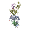
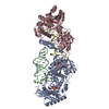
| Entry | Database: PDB / ID: 6nal | ||||||
|---|---|---|---|---|---|---|---|
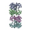
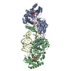
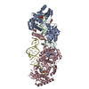
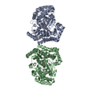
| Title | Crystal Structure of Gram Negative Toxin | ||||||
 Components Components | Thiol-activated cytolysin | ||||||
 Keywords Keywords | TOXIN / CYTOLYSIN | ||||||
| Function / homology |  Function and homology information Function and homology informationHIV-1 Reverse Transcriptase; Chain A, domain 3 / Thiol-activated cytolysin superfamily/Thiol-activated cytolysin, alpha-beta domain / Perfringolysin, domain 4 / Alpha-Beta Complex / Immunoglobulin-like / Sandwich / Mainly Beta / Alpha Beta Similarity search - Domain/homology | ||||||
| Biological species | Desulfobulbus propionicus | ||||||
| Method |  X-RAY DIFFRACTION / SYNCHROTRON / MAD / Resolution: 2.3 Å X-RAY DIFFRACTION / SYNCHROTRON / MAD / Resolution: 2.3 Å | ||||||
 Authors Authors | Morton, C.J. / Lawrence, S.A. / Parker, M.W. | ||||||
| Funding support |  Australia, 1items Australia, 1items
| ||||||
 Citation Citation | Journal: Mbio / Year: 2019 Title: The Structural Basis for a Transition State That Regulates Pore Formation in a Bacterial Toxin. Authors: Wade, K.R. / Lawrence, S.L. / Farrand, A.J. / Hotze, E.M. / Kuiper, M.J. / Gorman, M.A. / Christie, M.P. / Panjikar, S. / Morton, C.J. / Parker, M.W. / Tweten, R.K. | ||||||
| History |
|
- Structure visualization
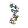
| Structure viewer | Molecule: MolmilJmol/JSmol |
|---|
- Downloads & links
-Download
| PDBx/mmCIF format | 6nal.cif.gz | 353.4 KB | Display | PDBx/mmCIF format |
|---|---|---|---|---|
| PDB format | pdb6nal.ent.gz | 303.4 KB | Display | PDB format |
| PDBx/mmJSON format | 6nal.json.gz | Tree view | PDBx/mmJSON format | |
| Others |  Other downloads Other downloads |
-Validation report
| Summary document | 6nal_validation.pdf.gz | 466.8 KB | Display | wwPDB validaton report |
|---|---|---|---|---|
| Full document | 6nal_full_validation.pdf.gz | 482.4 KB | Display | |
| Data in XML | 6nal_validation.xml.gz | 37.8 KB | Display | |
| Data in CIF | 6nal_validation.cif.gz | 52.8 KB | Display | |
| Arichive directory | https://data.pdbj.org/pub/pdb/validation_reports/na/6nalftp://data.pdbj.org/pub/pdb/validation_reports/na/6nal | HTTPS FTP |
-Related structure data
| Similar structure data |
|---|

-Links
 PDBj
PDBj


- Assembly
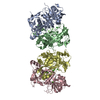
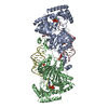
| Deposited unit | 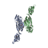
| ||||||||
|---|---|---|---|---|---|---|---|---|---|
| 1 | 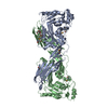
| ||||||||
| 2 | 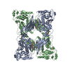
| ||||||||
| Unit cell |
|
-Components
| #1: Protein | Mass: 53010.605 Da / Num. of mol.: 2 Source method: isolated from a genetically manipulated source Source: (gene. exp.)  Desulfobulbus propionicus (strain ATCC 33891 / DSM 2032 / 1pr3) (bacteria) Desulfobulbus propionicus (strain ATCC 33891 / DSM 2032 / 1pr3) (bacteria)Strain: ATCC 33891 / DSM 2032 / 1pr3 / Gene: Despr_1128 / Production host: #2: Chemical | ChemComp-PG4 / |   Mass: 194.226 Da / Num. of mol.: 1 / Source method: obtained synthetically / Formula: C8H18O5 / Comment: precipitant*YM Mass: 194.226 Da / Num. of mol.: 1 / Source method: obtained synthetically / Formula: C8H18O5 / Comment: precipitant*YM#3: Chemical |   Mass: 69.085 Da / Num. of mol.: 2 / Source method: obtained synthetically / Formula: C3H5N2 Mass: 69.085 Da / Num. of mol.: 2 / Source method: obtained synthetically / Formula: C3H5N2#4: Water | ChemComp-HOH / |  Mass: 18.015 Da / Num. of mol.: 202 / Source method: isolated from a natural source / Formula: H2O Mass: 18.015 Da / Num. of mol.: 202 / Source method: isolated from a natural source / Formula: H2O |
|---|
-Experimental details
-Experiment
| Experiment | Method: X-RAY DIFFRACTION / Number of used crystals: 1 |
|---|
- Sample preparation
| Crystal | Density Matthews: 2.86 Å3/Da / Density % sol: 56.95 % |
|---|---|
| Crystal grow | Temperature: 294 K / Method: vapor diffusion, hanging drop / pH: 7.2 / Details: PEG 6000, 10% Tacsimate pH 5.0 |
-Data collection
| Diffraction | Mean temperature: 100 K / Serial crystal experiment: N | ||||||||||||||||||||||||
|---|---|---|---|---|---|---|---|---|---|---|---|---|---|---|---|---|---|---|---|---|---|---|---|---|---|
| Diffraction source | Source: SYNCHROTRON / Site: Australian Synchrotron / Beamline: MX2 / Wavelength: 0.95372, 0.97902, 0.97957 | ||||||||||||||||||||||||
| Detector | Type: DECTRIS EIGER X 16M / Detector: PIXEL / Date: Dec 9, 2016 | ||||||||||||||||||||||||
| Radiation | Protocol: MAD / Monochromatic (M) / Laue (L): M / Scattering type: x-ray | ||||||||||||||||||||||||
| Radiation wavelength |
| ||||||||||||||||||||||||
| Reflection | Resolution: 2.2→44.98 Å / Num. obs: 61001 / % possible obs: 100 % / Redundancy: 35.2 % / CC1/2: 0.997 / Rmerge(I) obs: 0.31 / Rpim(I) all: 0.049 / Rrim(I) all: 0.314 / Net I/σ(I): 11.4 / Num. measured all: 2147128 / Scaling rejects: 2403 | ||||||||||||||||||||||||
| Reflection shell | Diffraction-ID: 1
|
- Processing
| Software |
| ||||||||||||||||||||||||||||||||||||||||||||||||||||||||||||||||||||||||||||||||||||||||||||||||||||||||||||||||||||||||
|---|---|---|---|---|---|---|---|---|---|---|---|---|---|---|---|---|---|---|---|---|---|---|---|---|---|---|---|---|---|---|---|---|---|---|---|---|---|---|---|---|---|---|---|---|---|---|---|---|---|---|---|---|---|---|---|---|---|---|---|---|---|---|---|---|---|---|---|---|---|---|---|---|---|---|---|---|---|---|---|---|---|---|---|---|---|---|---|---|---|---|---|---|---|---|---|---|---|---|---|---|---|---|---|---|---|---|---|---|---|---|---|---|---|---|---|---|---|---|---|---|---|
| Refinement | Method to determine structure: MAD / Resolution: 2.3→43.445 Å / SU ML: 0.32 / Cross valid method: THROUGHOUT / σ(F): 1.34 / Phase error: 26.58
| ||||||||||||||||||||||||||||||||||||||||||||||||||||||||||||||||||||||||||||||||||||||||||||||||||||||||||||||||||||||||
| Solvent computation | Shrinkage radii: 0.9 Å / VDW probe radii: 1.11 Å | ||||||||||||||||||||||||||||||||||||||||||||||||||||||||||||||||||||||||||||||||||||||||||||||||||||||||||||||||||||||||
| Displacement parameters | Biso max: 204.49 Å2 / Biso mean: 59.2977 Å2 / Biso min: 22.74 Å2 | ||||||||||||||||||||||||||||||||||||||||||||||||||||||||||||||||||||||||||||||||||||||||||||||||||||||||||||||||||||||||
| Refinement step | Cycle: final / Resolution: 2.3→43.445 Å
| ||||||||||||||||||||||||||||||||||||||||||||||||||||||||||||||||||||||||||||||||||||||||||||||||||||||||||||||||||||||||
| LS refinement shell | Refine-ID: X-RAY DIFFRACTION / Rfactor Rfree error: 0 / Total num. of bins used: 19 / % reflection obs: 100 %
|
