 Movie
Movie Controller

[English] 日本語
 Yorodumi
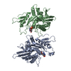
- PDB-6nag: X-ray structure of a secreted C11 cysteine protease from Bacteroi... -
+ Open data

- Basic information
| Entry | Database: PDB / ID: 6nag | ||||||
|---|---|---|---|---|---|---|---|
| Title | X-ray structure of a secreted C11 cysteine protease from Bacteroides thetaiotaomicron "iotapain | ||||||
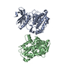
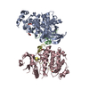
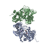
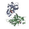
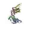
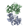
 Components Components | Clostripain-related protein | ||||||
 Keywords Keywords | HYDROLASE / C11 protease / secreted / microbiome / commensal | ||||||
| Function / homology | Peptidase C11, clostripain / Clostripain family / Prokaryotic membrane lipoprotein lipid attachment site profile. / PROLINE / Clostripain-related protein Function and homology information Function and homology information | ||||||
| Biological species |  Bacteroides thetaiotaomicron (bacteria) Bacteroides thetaiotaomicron (bacteria) | ||||||
| Method |  X-RAY DIFFRACTION / SYNCHROTRON / MOLECULAR REPLACEMENT / Resolution: 2.683 Å X-RAY DIFFRACTION / SYNCHROTRON / MOLECULAR REPLACEMENT / Resolution: 2.683 Å | ||||||
 Authors Authors | Wolan, D.W. / Gonzalez-Paez, G.E. / Roncase, E.J. | ||||||
 Citation Citation | Journal: Biochemistry / Year: 2019 Title: X-ray Structures of Two Bacteroides thetaiotaomicron C11 Proteases in Complex with Peptide-Based Inhibitors. Authors: Roncase, E.J. / Gonzalez-Paez, G.E. / Wolan, D.W. | ||||||
| History |
|
- Structure visualization
| Structure viewer | Molecule: MolmilJmol/JSmol |
|---|
- Downloads & links
-Download
| PDBx/mmCIF format | 6nag.cif.gz | 164.9 KB | Display | PDBx/mmCIF format |
|---|---|---|---|---|
| PDB format | pdb6nag.ent.gz | 128.2 KB | Display | PDB format |
| PDBx/mmJSON format | 6nag.json.gz | Tree view | PDBx/mmJSON format | |
| Others |  Other downloads Other downloads |
-Validation report
| Arichive directory | https://data.pdbj.org/pub/pdb/validation_reports/na/6nagftp://data.pdbj.org/pub/pdb/validation_reports/na/6nag | HTTPS FTP |
|---|
-Related structure data
| Related structure data | 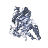 6n9jC 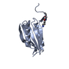 5l20S S: Starting model for refinement C: citing same article ( |
|---|---|
| Similar structure data |


-Links
 PDBj
PDBj


- Assembly
| Deposited unit | 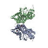
| ||||||||
|---|---|---|---|---|---|---|---|---|---|
| 1 |
| ||||||||
| Unit cell |
|
-Components
| #1: Protein | Mass: 41304.375 Da / Num. of mol.: 2 / Mutation: R154A Source method: isolated from a genetically manipulated source Source: (gene. exp.) Bacteroides thetaiotaomicron (strain ATCC 29148 / DSM 2079 / NCTC 10582 / E50 / VPI-5482) (bacteria)Strain: ATCC 29148 / DSM 2079 / NCTC 10582 / E50 / VPI-5482 / Gene: BT_0727 / Production host: #2: Chemical |   Type: L-peptide linking / Mass: 115.130 Da / Num. of mol.: 3 / Source method: obtained synthetically / Formula: C5H9NO2 / Details: Additive Type: L-peptide linking / Mass: 115.130 Da / Num. of mol.: 3 / Source method: obtained synthetically / Formula: C5H9NO2 / Details: Additive#3: Water | ChemComp-HOH / |  Mass: 18.015 Da / Num. of mol.: 607 / Source method: isolated from a natural source / Formula: H2O Mass: 18.015 Da / Num. of mol.: 607 / Source method: isolated from a natural source / Formula: H2O |
|---|
-Experimental details
-Experiment
| Experiment | Method: X-RAY DIFFRACTION / Number of used crystals: 1 |
|---|
- Sample preparation
| Crystal | Density Matthews: 4.8 Å3/Da / Density % sol: 74.38 % |
|---|---|
| Crystal grow | Temperature: 293 K / Method: vapor diffusion, sitting drop / pH: 5 / Details: 0.1 M Na Citrate, pH 5.0, 50% MPD, 10 mM L-Proline |
-Data collection
| Diffraction | Mean temperature: 100 K / Serial crystal experiment: N |
|---|---|
| Diffraction source | Source: SYNCHROTRON / Site: SSRL  / Beamline: BL9-2 / Wavelength: 0.9795 Å / Beamline: BL9-2 / Wavelength: 0.9795 Å |
| Detector | Type: DECTRIS PILATUS 6M / Detector: PIXEL / Date: Jan 19, 2017 / Details: Rh coated |
| Radiation | Monochromator: Si(111) / Protocol: SINGLE WAVELENGTH / Monochromatic (M) / Laue (L): M / Scattering type: x-ray |
| Radiation wavelength | Wavelength: 0.9795 Å / Relative weight: 1 |
| Reflection | Resolution: 2.68→45.52 Å / Num. obs: 47832 / % possible obs: 98.8 % / Redundancy: 4.6 % / Biso Wilson estimate: 29.8 Å2 / CC1/2: 0.986 / Rmerge(I) obs: 0.127 / Rpim(I) all: 0.081 / Rrim(I) all: 0.178 / Net I/σ(I): 11.2 |
| Reflection shell | Resolution: 2.68→2.73 Å / Redundancy: 4.5 % / Rmerge(I) obs: 0.58 / Mean I/σ(I) obs: 2.5 / Num. unique obs: 2368 / CC1/2: 0.719 / Rpim(I) all: 0.339 / Rrim(I) all: 0.735 / % possible all: 99 |
- Processing
| Software |
| |||||||||||||||||||||||||||||||||||||||||||||||||||||||||||||||
|---|---|---|---|---|---|---|---|---|---|---|---|---|---|---|---|---|---|---|---|---|---|---|---|---|---|---|---|---|---|---|---|---|---|---|---|---|---|---|---|---|---|---|---|---|---|---|---|---|---|---|---|---|---|---|---|---|---|---|---|---|---|---|---|---|
| Refinement | Method to determine structure: MOLECULAR REPLACEMENT Starting model: 5L20 Resolution: 2.683→45.517 Å / SU ML: 0.3 / Cross valid method: FREE R-VALUE / σ(F): 1.34 / Phase error: 19.93 / Stereochemistry target values: ML
| |||||||||||||||||||||||||||||||||||||||||||||||||||||||||||||||
| Solvent computation | Shrinkage radii: 0.9 Å / VDW probe radii: 1.11 Å / Solvent model: FLAT BULK SOLVENT MODEL | |||||||||||||||||||||||||||||||||||||||||||||||||||||||||||||||
| Refinement step | Cycle: LAST / Resolution: 2.683→45.517 Å
| |||||||||||||||||||||||||||||||||||||||||||||||||||||||||||||||
| Refine LS restraints |
| |||||||||||||||||||||||||||||||||||||||||||||||||||||||||||||||
| LS refinement shell |
|
