 Movie
Movie Controller


+ Open data

- Basic information
| Entry | Database: PDB / ID: 6n8v | ||||||
|---|---|---|---|---|---|---|---|
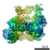
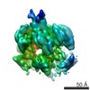
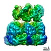
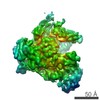
| Title | Hsp104DWB open conformation | ||||||
 Components Components | Heat shock protein 104 | ||||||
 Keywords Keywords | CHAPERONE / Hsp104 / ClpB / protein disaggregase / molecular chaperone / AAA+ / ATPase / cryo-EM | ||||||
| Function / homology |  Function and homology information Function and homology informationtrehalose metabolic process / TRC complex / protein folding in endoplasmic reticulum / cellular heat acclimation / post-translational protein targeting to endoplasmic reticulum membrane / stress granule disassembly / : / protein unfolding / nuclear periphery / ADP binding ...trehalose metabolic process / TRC complex / protein folding in endoplasmic reticulum / cellular heat acclimation / post-translational protein targeting to endoplasmic reticulum membrane / stress granule disassembly / : / protein unfolding / nuclear periphery / ADP binding / unfolded protein binding / protein-folding chaperone binding / cellular response to heat / protein refolding / ATP hydrolysis activity / ATP binding / identical protein binding / nucleus / cytosol / cytoplasm Similarity search - Function | ||||||
| Biological species |  | ||||||
| Method | ELECTRON MICROSCOPY / single particle reconstruction / cryo EM / Resolution: 9.3 Å | ||||||
 Authors Authors | Lee, S. / Rho, S.H. / Lee, J. / Sung, N. / Liu, J. / Tsai, F.T.F. | ||||||
 Citation Citation | Journal: Cell Rep / Year: 2019 Title: Cryo-EM Structures of the Hsp104 Protein Disaggregase Captured in the ATP Conformation. Authors: Sukyeong Lee / Soung Hun Roh / Jungsoon Lee / Nuri Sung / Jun Liu / Francis T F Tsai /  Abstract: Hsp104 is a ring-forming, ATP-driven molecular machine that recovers functional protein from both stress-denatured and amyloid-forming aggregates. Although Hsp104 shares a common architecture with ...Hsp104 is a ring-forming, ATP-driven molecular machine that recovers functional protein from both stress-denatured and amyloid-forming aggregates. Although Hsp104 shares a common architecture with Clp/Hsp100 protein unfoldases, different and seemingly conflicting 3D structures have been reported. Examining the structure of Hsp104 poses considerable challenges because Hsp104 readily hydrolyzes ATP, whereas ATP analogs can be slowly turned over and are often contaminated with other nucleotide species. Here, we present the single-particle electron cryo-microscopy (cryo-EM) structures of a catalytically inactive Hsp104 variant (Hsp104) in the ATP-bound state determined between 7.7 Å and 9.3 Å resolution. Surprisingly, we observe that the Hsp104 hexamer adopts distinct ring conformations (closed, extended, and open) despite being in the same nucleotide state. The latter underscores the structural plasticity of Hsp104 in solution, with different conformations stabilized by nucleotide binding. Our findings suggest that, in addition to ATP hydrolysis-driven conformational changes, Hsp104 uses stochastic motions to translocate unfolded polypeptides. | ||||||
| History |
|
- Structure visualization
| Movie |
Movie viewer |
|---|---|
| Structure viewer | Molecule: MolmilJmol/JSmol |
- Downloads & links
-Download
| PDBx/mmCIF format | 6n8v.cif.gz | 566.1 KB | Display | PDBx/mmCIF format |
|---|---|---|---|---|
| PDB format | pdb6n8v.ent.gz | 347.1 KB | Display | PDB format |
| PDBx/mmJSON format | 6n8v.json.gz | Tree view | PDBx/mmJSON format | |
| Others |  Other downloads Other downloads |
-Validation report
| Summary document | 6n8v_validation.pdf.gz | 1.6 MB | Display | wwPDB validaton report |
|---|---|---|---|---|
| Full document | 6n8v_full_validation.pdf.gz | 1.7 MB | Display | |
| Data in XML | 6n8v_validation.xml.gz | 104.8 KB | Display | |
| Data in CIF | 6n8v_validation.cif.gz | 167 KB | Display | |
| Arichive directory | https://data.pdbj.org/pub/pdb/validation_reports/n8/6n8vftp://data.pdbj.org/pub/pdb/validation_reports/n8/6n8v | HTTPS FTP |
-Related structure data
| Related structure data |  0376MC 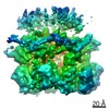 0375C 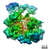 0377C 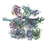 6n8tC 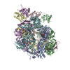 6n8zC C: citing same article ( M: map data used to model this data |
|---|---|
| Similar structure data |

-Links
 PDBj
PDBj


- Assembly
| Deposited unit | 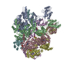
|
|---|---|
| 1 |
|
-Components
| #1: Protein | Mass: 99046.859 Da / Num. of mol.: 6 Source method: isolated from a genetically manipulated source Source: (gene. exp.) Gene: HSP104, YLL026W, L0948 / Production host:  #2: Chemical | ChemComp-ATP /   Mass: 507.181 Da / Num. of mol.: 11 / Source method: obtained synthetically / Formula: C10H16N5O13P3 / Comment: ATP, energy-carrying molecule*YM Mass: 507.181 Da / Num. of mol.: 11 / Source method: obtained synthetically / Formula: C10H16N5O13P3 / Comment: ATP, energy-carrying molecule*YM |
|---|
-Experimental details
-Experiment
| Experiment | Method: ELECTRON MICROSCOPY |
|---|---|
| EM experiment | Aggregation state: PARTICLE / 3D reconstruction method: single particle reconstruction |
- Sample preparation
| Component | Name: Hexamer of Hsp104 with ATP / Type: COMPLEX / Details: Hsp 104 double Walker B mutant / Entity ID: #1 / Source: RECOMBINANT | ||||||||||||||||||||
|---|---|---|---|---|---|---|---|---|---|---|---|---|---|---|---|---|---|---|---|---|---|
| Molecular weight | Value: 0.6 MDa / Experimental value: YES | ||||||||||||||||||||
| Source (natural) | Organism: | ||||||||||||||||||||
| Source (recombinant) | Organism: | ||||||||||||||||||||
| Buffer solution | pH: 7.5 / Details: Buffer solution is freshly prepared. | ||||||||||||||||||||
| Buffer component |
| ||||||||||||||||||||
| Specimen | Conc.: 0.3 mg/ml / Embedding applied: NO / Shadowing applied: NO / Staining applied: NO / Vitrification applied: YES / Details: This sample was mono disperse. | ||||||||||||||||||||
| Specimen support | Details: unspecified | ||||||||||||||||||||
| Vitrification | Instrument: FEI VITROBOT MARK IV / Cryogen name: ETHANE / Humidity: 100 % / Chamber temperature: 297 K / Details: blot for 3 seconds before plunging |
- Electron microscopy imaging
| Experimental equipment |  Model: Tecnai Polara / Image courtesy: FEI Company |
|---|---|
| Microscopy | Model: FEI POLARA 300 |
| Electron gun | Electron source:  FIELD EMISSION GUN / Accelerating voltage: 300 kV / Illumination mode: FLOOD BEAM FIELD EMISSION GUN / Accelerating voltage: 300 kV / Illumination mode: FLOOD BEAM |
| Electron lens | Mode: BRIGHT FIELD |
| Image recording | Average exposure time: 7.6 sec. / Electron dose: 38 e/Å2 / Detector mode: SUPER-RESOLUTION / Film or detector model: GATAN K2 SUMMIT (4k x 4k) / Num. of real images: 1394 |
- Processing
| CTF correction | Type: PHASE FLIPPING AND AMPLITUDE CORRECTION |
|---|---|
| 3D reconstruction | Resolution: 9.3 Å / Resolution method: FSC 0.143 CUT-OFF / Num. of particles: 25582 / Symmetry type: POINT |
| Refinement | Highest resolution: 9.3 Å |
