 Movie
Movie Controller


+ Open data

- Basic information
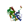
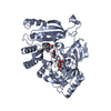
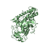
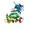
| Entry | Database: PDB / ID: 6n84 | ||||||||||||
|---|---|---|---|---|---|---|---|---|---|---|---|---|---|
| Title | MBP-fusion protein of transducin-alpha residues 327-350 | ||||||||||||
 Components Components | Maltose/maltodextrin-binding periplasmic protein,Guanine nucleotide-binding protein G(t) subunit alpha-2 | ||||||||||||
 Keywords Keywords | CHAPERONE / G alpha / MBP | ||||||||||||
| Function / homology |  Function and homology information Function and homology informationG protein-coupled photoreceptor activity / detection of light stimulus involved in visual perception / response to light intensity / detection of chemical stimulus involved in sensory perception of bitter taste / photoreceptor outer segment membrane / detection of maltose stimulus / maltose transport complex / carbohydrate transport / phototransduction / carbohydrate transmembrane transporter activity ...G protein-coupled photoreceptor activity / detection of light stimulus involved in visual perception / response to light intensity / detection of chemical stimulus involved in sensory perception of bitter taste / photoreceptor outer segment membrane / detection of maltose stimulus / maltose transport complex / carbohydrate transport / phototransduction / carbohydrate transmembrane transporter activity / photoreceptor outer segment / maltose binding / maltose transport / maltodextrin transmembrane transport / ATP-binding cassette (ABC) transporter complex, substrate-binding subunit-containing / photoreceptor inner segment / visual perception / ATP-binding cassette (ABC) transporter complex / cell chemotaxis / G protein-coupled receptor binding / G-protein beta/gamma-subunit complex binding / adenylate cyclase-modulating G protein-coupled receptor signaling pathway / outer membrane-bounded periplasmic space / heterotrimeric G-protein complex / positive regulation of cytosolic calcium ion concentration / Ca2+ pathway / G alpha (i) signalling events / periplasmic space / G protein-coupled receptor signaling pathway / GTPase activity / DNA damage response / synapse / GTP binding / membrane / metal ion binding / plasma membrane / cytoplasm Similarity search - Function | ||||||||||||
| Biological species |   Homo sapiens (human) Homo sapiens (human) | ||||||||||||
| Method |  X-RAY DIFFRACTION / SYNCHROTRON / MOLECULAR REPLACEMENT / Resolution: 1.75 Å X-RAY DIFFRACTION / SYNCHROTRON / MOLECULAR REPLACEMENT / Resolution: 1.75 Å | ||||||||||||
 Authors Authors | Srivastava, D. / Gakhar, L. / Artemyev, N.O. | ||||||||||||
| Funding support |  United States, 1items United States, 1items
| ||||||||||||
 Citation Citation | Journal: Nat Commun / Year: 2019 Title: Structural underpinnings of Ric8A function as a G-protein α-subunit chaperone and guanine-nucleotide exchange factor. Authors: Dhiraj Srivastava / Lokesh Gakhar / Nikolai O Artemyev / Abstract: Resistance to inhibitors of cholinesterase 8A (Ric8A) is an essential regulator of G protein α-subunits (Gα), acting as a guanine nucleotide exchange factor and a chaperone. We report two crystal ...Resistance to inhibitors of cholinesterase 8A (Ric8A) is an essential regulator of G protein α-subunits (Gα), acting as a guanine nucleotide exchange factor and a chaperone. We report two crystal structures of Ric8A, one in the apo form and the other in complex with a tagged C-terminal fragment of Gα. These structures reveal two principal domains of Ric8A: an armadillo-fold core and a flexible C-terminal tail. Additionally, they show that the Gα C-terminus binds to a highly-conserved patch on the concave surface of the Ric8A armadillo-domain, with selectivity determinants residing in the Gα sequence. Biochemical analysis shows that the Ric8A C-terminal tail is critical for its stability and function. A model of the Ric8A/Gα complex derived from crosslinking mass spectrometry and molecular dynamics simulations suggests that the Ric8A C-terminal tail helps organize the GTP-binding site of Gα. This study lays the groundwork for understanding Ric8A function at the molecular level. | ||||||||||||
| History |
|
- Structure visualization
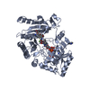
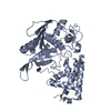
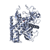
| Structure viewer | Molecule: MolmilJmol/JSmol |
|---|
- Downloads & links
-Download
| PDBx/mmCIF format | 6n84.cif.gz | 191.4 KB | Display | PDBx/mmCIF format |
|---|---|---|---|---|
| PDB format | pdb6n84.ent.gz | 148.5 KB | Display | PDB format |
| PDBx/mmJSON format | 6n84.json.gz | Tree view | PDBx/mmJSON format | |
| Others |  Other downloads Other downloads |
-Validation report
| Arichive directory | https://data.pdbj.org/pub/pdb/validation_reports/n8/6n84ftp://data.pdbj.org/pub/pdb/validation_reports/n8/6n84 | HTTPS FTP |
|---|
-Related structure data
| Related structure data |  6n85C  6n86C  1anfS S: Starting model for refinement C: citing same article ( |
|---|---|
| Similar structure data |


-Links
 PDBj
PDBj


- Assembly
| Deposited unit | 
| ||||||||||||||||||
|---|---|---|---|---|---|---|---|---|---|---|---|---|---|---|---|---|---|---|---|
| 1 |
| ||||||||||||||||||
| Unit cell |
| ||||||||||||||||||
| Components on special symmetry positions |
|
-Components
| #1: Protein | Mass: 45448.391 Da / Num. of mol.: 1 / Mutation: D83A, K84A, E173A, N174A, A216H, K220H, K240A Source method: isolated from a genetically manipulated source Source: (gene. exp.) Homo sapiens (human)Gene: malE, Z5632, ECs5017, GNAT2, GNATC / Production host: References: UniProt: P0AEY0, UniProt: P19087, UniProt: P0AEX9*PLUS | ||
|---|---|---|---|
| #2: Polysaccharide | alpha-D-glucopyranose-(1-4)-alpha-D-glucopyranose / alpha-maltose  Source method: isolated from a genetically manipulated source Details: oligosaccharide / References: alpha-maltose | ||
| #3: Chemical | ChemComp-SO4 /   Mass: 96.063 Da / Num. of mol.: 8 Mass: 96.063 Da / Num. of mol.: 8Source method: isolated from a genetically manipulated source Formula: SO4 #4: Water | ChemComp-HOH / |  Mass: 18.015 Da / Num. of mol.: 478 / Source method: isolated from a natural source / Formula: H2O Mass: 18.015 Da / Num. of mol.: 478 / Source method: isolated from a natural source / Formula: H2O |
-Experimental details
-Experiment
| Experiment | Method: X-RAY DIFFRACTION / Number of used crystals: 1 |
|---|
- Sample preparation
| Crystal | Density Matthews: 2.39 Å3/Da / Density % sol: 48.55 % |
|---|---|
| Crystal grow | Temperature: 291 K / Method: vapor diffusion, hanging drop / pH: 4.6 Details: 2 M Ammonium Sulphate, 0.1 M sodium acetate, pH 4.6 |
-Data collection
| Diffraction | Mean temperature: 100 K / Serial crystal experiment: N |
|---|---|
| Diffraction source | Source: SYNCHROTRON / Site: ALS / Beamline: 4.2.2 / Wavelength: 1 Å |
| Detector | Type: RDI CMOS_8M / Detector: CMOS / Date: Oct 10, 2017 |
| Radiation | Protocol: SINGLE WAVELENGTH / Monochromatic (M) / Laue (L): M / Scattering type: x-ray |
| Radiation wavelength | Wavelength: 1 Å / Relative weight: 1 |
| Reflection | Resolution: 1.75→60.08 Å / Num. obs: 43096 / % possible obs: 100 % / Redundancy: 10.7 % / CC1/2: 1 / Rmerge(I) obs: 0.062 / Rpim(I) all: 0.021 / Rrim(I) all: 0.069 / Net I/σ(I): 29 |
| Reflection shell | Resolution: 1.75→1.84 Å / Redundancy: 9.3 % / Rmerge(I) obs: 0.753 / Mean I/σ(I) obs: 2.5 / Num. unique obs: 6311 / CC1/2: 0.809 / Rpim(I) all: 0.277 / Rrim(I) all: 0.849 / % possible all: 100 |
- Processing
| Software |
| ||||||||||||||||||||||||||||||||||||||||||||||||||||||||||||||||||||||||||||||||||||||||||||||||||||||||||||||||
|---|---|---|---|---|---|---|---|---|---|---|---|---|---|---|---|---|---|---|---|---|---|---|---|---|---|---|---|---|---|---|---|---|---|---|---|---|---|---|---|---|---|---|---|---|---|---|---|---|---|---|---|---|---|---|---|---|---|---|---|---|---|---|---|---|---|---|---|---|---|---|---|---|---|---|---|---|---|---|---|---|---|---|---|---|---|---|---|---|---|---|---|---|---|---|---|---|---|---|---|---|---|---|---|---|---|---|---|---|---|---|---|---|---|
| Refinement | Method to determine structure: MOLECULAR REPLACEMENT Starting model: 1ANF Resolution: 1.75→60.08 Å / SU ML: 0.16 / Cross valid method: FREE R-VALUE / σ(F): 1.35 / Phase error: 18.6 / Stereochemistry target values: ML
| ||||||||||||||||||||||||||||||||||||||||||||||||||||||||||||||||||||||||||||||||||||||||||||||||||||||||||||||||
| Solvent computation | Shrinkage radii: 0.9 Å / VDW probe radii: 1.11 Å / Solvent model: FLAT BULK SOLVENT MODEL | ||||||||||||||||||||||||||||||||||||||||||||||||||||||||||||||||||||||||||||||||||||||||||||||||||||||||||||||||
| Refinement step | Cycle: LAST / Resolution: 1.75→60.08 Å
| ||||||||||||||||||||||||||||||||||||||||||||||||||||||||||||||||||||||||||||||||||||||||||||||||||||||||||||||||
| Refine LS restraints |
| ||||||||||||||||||||||||||||||||||||||||||||||||||||||||||||||||||||||||||||||||||||||||||||||||||||||||||||||||
| LS refinement shell |
| ||||||||||||||||||||||||||||||||||||||||||||||||||||||||||||||||||||||||||||||||||||||||||||||||||||||||||||||||
| Refinement TLS params. | Method: refined / Origin x: 99.8971 Å / Origin y: 25.7937 Å / Origin z: 2.6083 Å
| ||||||||||||||||||||||||||||||||||||||||||||||||||||||||||||||||||||||||||||||||||||||||||||||||||||||||||||||||
| Refinement TLS group | Selection details: all |
