 Movie
Movie Controller

[English] 日本語
 Yorodumi
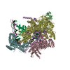
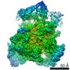
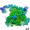
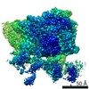
- PDB-6n4c: EM structure of the DNA wrapping in bacterial open transcription ... -
+ Open data

- Basic information
| Entry | Database: PDB / ID: 6n4c | ||||||
|---|---|---|---|---|---|---|---|
| Title | EM structure of the DNA wrapping in bacterial open transcription initiation complex | ||||||
 Components Components |
| ||||||
 Keywords Keywords | transcription/dna / DNA wrapping / bacterial transcription initiation complex / transmission electron microscopy / single particle analysis. / TRANSCRIPTION / transcription-dna complex | ||||||
| Function / homology |  Function and homology information Function and homology informationsigma factor antagonist complex / RNA polymerase complex / submerged biofilm formation / cellular response to cell envelope stress / regulation of DNA-templated transcription initiation / sigma factor activity / bacterial-type flagellum assembly / bacterial-type RNA polymerase core enzyme binding / cytosolic DNA-directed RNA polymerase complex / bacterial-type flagellum-dependent cell motility ...sigma factor antagonist complex / RNA polymerase complex / submerged biofilm formation / cellular response to cell envelope stress / regulation of DNA-templated transcription initiation / sigma factor activity / bacterial-type flagellum assembly / bacterial-type RNA polymerase core enzyme binding / cytosolic DNA-directed RNA polymerase complex / bacterial-type flagellum-dependent cell motility / nitrate assimilation / regulation of DNA-templated transcription elongation / transcription elongation factor complex / DNA-directed RNA polymerase complex / transcription antitermination / DNA-templated transcription initiation / cell motility / ribonucleoside binding / DNA-directed RNA polymerase / DNA-directed RNA polymerase activity / response to heat / protein-containing complex assembly / intracellular iron ion homeostasis / protein dimerization activity / transcription cis-regulatory region binding / response to antibiotic / negative regulation of DNA-templated transcription / regulation of DNA-templated transcription / DNA-templated transcription / magnesium ion binding / DNA binding / zinc ion binding / membrane / cytoplasm / cytosol Similarity search - Function | ||||||
| Biological species |   Enterobacteria phage lambda (virus) Enterobacteria phage lambda (virus) | ||||||
| Method | ELECTRON MICROSCOPY / single particle reconstruction / negative staining / Resolution: 17 Å | ||||||
 Authors Authors | Florez-Ariza, A. / Cassago, A. / de Oliveira, P.S.L. / Guerra, D.G. / Portugal, R.V. | ||||||
 Citation Citation | Journal: Biorxiv / Year: 2020 Title: Interactions of Upstream and Downstream Promoter Regions with RNA Polymerase are Energetically Coupled and a Target of Regulation in Transcription Initiation Authors: Sosa, R. / Florez-Ariza, A. / Diaz-Celis, C. / Onoa, B. / Cassago, A. / de Oliveira, P.S.L. / Portugal, R.V. / Guerra, D.G. / Bustamante, C. | ||||||
| History |
|
- Structure visualization
| Movie |
Movie viewer |
|---|---|
| Structure viewer | Molecule: MolmilJmol/JSmol |
 UCSF Chimera
- Downloads & links
-Download
| PDBx/mmCIF format | 6n4c.cif.gz | 1.5 MB | Display | PDBx/mmCIF format |
|---|---|---|---|---|
| PDB format | pdb6n4c.ent.gz | 1.3 MB | Display | PDB format |
| PDBx/mmJSON format | 6n4c.json.gz | Tree view | PDBx/mmJSON format | |
| Others |  Other downloads Other downloads |
-Validation report
| Arichive directory | https://data.pdbj.org/pub/pdb/validation_reports/n4/6n4cftp://data.pdbj.org/pub/pdb/validation_reports/n4/6n4c | HTTPS FTP |
|---|
-Related structure data
| Related structure data |  0340MC M: map data used to model this data C: citing same article ( |
|---|---|
| Similar structure data |


-Links
 PDBj
PDBj


- Assembly
| Deposited unit | 
|
|---|---|
| 1 |
|
-Components
-Protein , 1 types, 1 molecules F
| #1: Protein | Mass: 64322.277 Da / Num. of mol.: 1 / Source method: isolated from a natural source / Source: (natural) |
|---|
-DNA-directed RNA polymerase subunit ... , 5 types, 5 molecules CDABE
| #2: Protein | Mass: 150689.672 Da / Num. of mol.: 1 / Source method: isolated from a natural source / Source: (natural) References: UniProt: A0A0A0GWV9, UniProt: P0A8V2*PLUS, DNA-directed RNA polymerase |
|---|---|
| #3: Protein | Mass: 150320.219 Da / Num. of mol.: 1 / Source method: isolated from a natural source / Source: (natural) References: UniProt: A0A369F490, UniProt: P0A8T7*PLUS, DNA-directed RNA polymerase |
| #4: Protein | Mass: 35275.273 Da / Num. of mol.: 1 / Source method: isolated from a natural source / Source: (natural) |
| #5: Protein | Mass: 34400.266 Da / Num. of mol.: 1 / Source method: isolated from a natural source / Source: (natural) |
| #6: Protein | Mass: 10118.352 Da / Num. of mol.: 1 / Source method: isolated from a natural source / Source: (natural) |
-DNA chain , 2 types, 2 molecules ab
| #7: DNA chain | Mass: 29094.617 Da / Num. of mol.: 1 / Source method: obtained synthetically / Details: NT strand DNA (94-MER) / Source: (synth.) Enterobacteria phage lambda (virus) |
|---|---|
| #8: DNA chain | Mass: 28890.529 Da / Num. of mol.: 1 / Source method: obtained synthetically / Details: T strand DNA (94-MER) / Source: (synth.) Enterobacteria phage lambda (virus) |
-Details
| Has protein modification | Y |
|---|
-Experimental details
-Experiment
| Experiment | Method: ELECTRON MICROSCOPY |
|---|---|
| EM experiment | Aggregation state: PARTICLE / 3D reconstruction method: single particle reconstruction |
- Sample preparation
| Component | Name: E. coli RNAP-DNA wrapped open complex / Type: COMPLEX Details: Wrapped transcription initiation open complex assembled between E. coli RNAP-sigma 70 holoenzyme and lambda PR wild-type promoter (+18 to -76) Entity ID: all / Source: MULTIPLE SOURCES |
|---|---|
| Molecular weight | Value: 0.50 MDa / Experimental value: NO |
| Source (natural) | Organism: |
| Buffer solution | pH: 7.9 |
| Specimen | Embedding applied: NO / Shadowing applied: NO / Staining applied: YES / Vitrification applied: NO |
| EM staining | Type: NEGATIVE Details: Negatively stained EM specimens were prepared using a 2% uranyl acetate solution. Material: Uranyl Acetate |
| Specimen support | Details: na |
- Electron microscopy imaging
| Microscopy | Model: JEOL 2100 |
|---|---|
| Electron gun | Electron source: LAB6 / Accelerating voltage: 200 kV / Illumination mode: FLOOD BEAM |
| Electron lens | Mode: BRIGHT FIELD |
| Image recording | Electron dose: 20 e/Å2 / Film or detector model: TVIPS TEMCAM-F416 (4k x 4k) |
- Processing
| EM software |
| |||||||||||||||||||||
|---|---|---|---|---|---|---|---|---|---|---|---|---|---|---|---|---|---|---|---|---|---|---|
| CTF correction | Type: PHASE FLIPPING ONLY | |||||||||||||||||||||
| Particle selection | Num. of particles selected: 60393 | |||||||||||||||||||||
| Symmetry | Point symmetry: C1 (asymmetric) | |||||||||||||||||||||
| 3D reconstruction | Resolution: 17 Å / Resolution method: FSC 1/2 BIT CUT-OFF / Num. of particles: 16015 / Symmetry type: POINT | |||||||||||||||||||||
| Atomic model building | Protocol: OTHER Details: Initial fiting was done using manual fitting in Chimera. Yasara software was used for energy minimizaton of the DNA and RNAP coordinates model. | |||||||||||||||||||||
| Atomic model building | PDB-ID: 4YLN Accession code: 4YLN / Source name: PDB / Type: experimental model |

