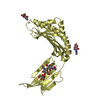
endopeptidase La / ATP-dependent peptidase activity / protein quality control for misfolded or incompletely synthesized proteins / response to X-ray / peptidase activity / cellular response to heat / response to heat / sequence-specific DNA binding / serine-type endopeptidase activity / ATP hydrolysis activity ...endopeptidase La / ATP-dependent peptidase activity / protein quality control for misfolded or incompletely synthesized proteins / response to X-ray / peptidase activity / cellular response to heat / response to heat / sequence-specific DNA binding / serine-type endopeptidase activity / ATP hydrolysis activity / proteolysis / DNA binding / ATP binding / cytoplasm / cytosol Similarity search - Function
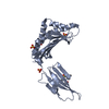
Resolution: 3.5→44.3 Å / SU ML: 0.45 / Cross valid method: FREE R-VALUE / σ(F): 1.34 / Phase error: 29.93 Details: While the interactions are not very precise at atomic level, the structure still provides valuable information about the quaternary state of the molecule.
Rfactor
Num. reflection
% reflection
Rfree
0.3254
219
5.68 %
Rwork
0.2845
-
-
obs
0.2866
3856
99 %
Solvent computation
Shrinkage radii: 0.9 Å / VDW probe radii: 1.11 Å
Refinement step
Cycle: LAST / Resolution: 3.5→44.3 Å
Protein
Nucleic acid
Ligand
Solvent
Total
Num. atoms
2669
0
27
0
2696
Refine LS restraints
Refine-ID
Type
Dev ideal
Number
X-RAY DIFFRACTION
f_bond_d
0.003
2736
X-RAY DIFFRACTION
f_angle_d
0.661
3683
X-RAY DIFFRACTION
f_dihedral_angle_d
13.704
1716
X-RAY DIFFRACTION
f_chiral_restr
0.041
412
X-RAY DIFFRACTION
f_plane_restr
0.004
471
LS refinement shell
Resolution (Å)
Rfactor Rfree
Num. reflection Rfree
Rfactor Rwork
Num. reflection Rwork
Refine-ID
% reflection obs (%)
3.5002-4.4093
0.3893
119
0.3388
1776
X-RAY DIFFRACTION
98
4.4093-44.3035
0.2844
100
0.2555
1861
X-RAY DIFFRACTION
100
+
About Yorodumi
-
News
-
Feb 9, 2022. New format data for meta-information of EMDB entries
New format data for meta-information of EMDB entries
Version 3 of the EMDB header file is now the official format.
The previous official version 1.9 will be removed from the archive.
In the structure databanks used in Yorodumi, some data are registered as the other names, "COVID-19 virus" and "2019-nCoV". Here are the details of the virus and the list of structure data.
Jan 31, 2019. EMDB accession codes are about to change! (news from PDBe EMDB page)
EMDB accession codes are about to change! (news from PDBe EMDB page)
The allocation of 4 digits for EMDB accession codes will soon come to an end. Whilst these codes will remain in use, new EMDB accession codes will include an additional digit and will expand incrementally as the available range of codes is exhausted. The current 4-digit format prefixed with “EMD-” (i.e. EMD-XXXX) will advance to a 5-digit format (i.e. EMD-XXXXX), and so on. It is currently estimated that the 4-digit codes will be depleted around Spring 2019, at which point the 5-digit format will come into force.
The EM Navigator/Yorodumi systems omit the EMD- prefix.
Related info.:Q: What is EMD? / ID/Accession-code notation in Yorodumi/EM Navigator
Yorodumi is a browser for structure data from EMDB, PDB, SASBDB, etc.
This page is also the successor to EM Navigator detail page, and also detail information page/front-end page for Omokage search.
The word "yorodu" (or yorozu) is an old Japanese word meaning "ten thousand". "mi" (miru) is to see.
Related info.:EMDB / PDB / SASBDB / Comparison of 3 databanks / Yorodumi Search / Aug 31, 2016. New EM Navigator & Yorodumi / Yorodumi Papers / Jmol/JSmol / Function and homology information / Changes in new EM Navigator and Yorodumi
 Movie
Movie Controller


 Open data
 Basic information
 Components
 Keywords
 Function and homology information
 X-RAY DIFFRACTION /
  Authors
 United States, 1items
  Citation
 Structure visualization
 Downloads & links
 Other downloads
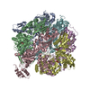


 PDBj
PDBj
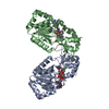
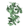
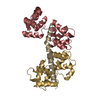
 Assembly

 Mass: 427.201 Da / Num. of mol.: 1 / Source method: obtained synthetically / Formula: C10H15N5O10P2 / Comment: ADP, energy-carrying molecule*YM
 Sample preparation
 Processing