 Movie
Movie Controller

[English] 日本語
 Yorodumi
- PDB-6muk: 1.93 Angstrom Resolution Crystal Structure of Peptidase M23 from ... -
+ Open data

- Basic information
| Entry | Database: PDB / ID: 6muk | |||||||||
|---|---|---|---|---|---|---|---|---|---|---|
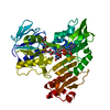
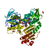
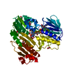
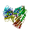
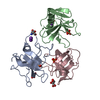
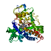
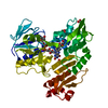
| Title | 1.93 Angstrom Resolution Crystal Structure of Peptidase M23 from Neisseria gonorrhoeae. | |||||||||
 Components Components | Peptidase M23 | |||||||||
 Keywords Keywords | HYDROLASE / Structural Genomics / Center for Structural Genomics of Infectious Diseases / CSGID / Peptidase M23 | |||||||||
| Function / homology | NMB0315-like, domain I / : / Peptidase M23 / Peptidase family M23 / Duplicated hybrid motif / metal ion binding / Peptidase M23 Function and homology information Function and homology information | |||||||||
| Biological species |  Neisseria gonorrhoeae (bacteria) Neisseria gonorrhoeae (bacteria) | |||||||||
| Method |  X-RAY DIFFRACTION / SYNCHROTRON / MOLECULAR REPLACEMENT / Resolution: 1.93 Å X-RAY DIFFRACTION / SYNCHROTRON / MOLECULAR REPLACEMENT / Resolution: 1.93 Å | |||||||||
 Authors Authors | Minasov, G. / Shuvalova, L. / Pshenychnyi, S. / Satchell, K.J.F. / Joachimiak, A. / Center for Structural Genomics of Infectious Diseases (CSGID) | |||||||||
 Citation Citation | Journal: To Be Published Title: 1.93 Angstrom Resolution Crystal Structure of Peptidase M23 from Neisseria gonorrhoeae. Authors: Minasov, G. / Shuvalova, L. / Pshenychnyi, S. / Satchell, K.J.F. / Joachimiak, A. / Center for Structural Genomics of Infectious Diseases (CSGID) | |||||||||
| History |
|
- Structure visualization
| Structure viewer | Molecule: MolmilJmol/JSmol |
|---|
- Downloads & links
-Download
| PDBx/mmCIF format | 6muk.cif.gz | 165.3 KB | Display | PDBx/mmCIF format |
|---|---|---|---|---|
| PDB format | pdb6muk.ent.gz | 129.3 KB | Display | PDB format |
| PDBx/mmJSON format | 6muk.json.gz | Tree view | PDBx/mmJSON format | |
| Others |  Other downloads Other downloads |
-Validation report
| Arichive directory | https://data.pdbj.org/pub/pdb/validation_reports/mu/6mukftp://data.pdbj.org/pub/pdb/validation_reports/mu/6muk | HTTPS FTP |
|---|
-Related structure data
| Related structure data |  3sluS S: Starting model for refinement |
|---|---|
| Similar structure data | |
| Other databases |
-Links
 PDBj
PDBj- Assembly
| Deposited unit | 
| ||||||||
|---|---|---|---|---|---|---|---|---|---|
| 1 |
| ||||||||
| Unit cell |
|
-Components
| #1: Protein | Mass: 43199.203 Da / Num. of mol.: 1 Source method: isolated from a genetically manipulated source Source: (gene. exp.) Neisseria gonorrhoeae (bacteria) / Strain: ATCC 700825 / FA 1090 / Gene: NGO_1686 / Plasmid: pET28aProduction host: Strain (production host): BL21.Gold / References: UniProt: Q5F676 |
|---|---|
| #2: Chemical | ChemComp-ZN /   Mass: 65.409 Da / Num. of mol.: 1 / Source method: obtained synthetically / Formula: Zn Mass: 65.409 Da / Num. of mol.: 1 / Source method: obtained synthetically / Formula: Zn |
| #3: Water | ChemComp-HOH /  Mass: 18.015 Da / Num. of mol.: 222 / Source method: isolated from a natural source / Formula: H2O Mass: 18.015 Da / Num. of mol.: 222 / Source method: isolated from a natural source / Formula: H2O |
-Experimental details
-Experiment
| Experiment | Method: X-RAY DIFFRACTION / Number of used crystals: 1 |
|---|
- Sample preparation
| Crystal | Density Matthews: 2.26 Å3/Da / Density % sol: 45.7 % |
|---|---|
| Crystal grow | Temperature: 292 K / Method: vapor diffusion, sitting drop / pH: 7 Details: Protein: 13.0 mg/ml, 0.5M Sodium chloride, 0.01M Tris-HCl pH 8.3; Screen: Classics II (C2), 1.1M Ammonium tartrate pH 7.0; Cryo: 4M Sodium formate. |
-Data collection
| Diffraction | Mean temperature: 100 K / Serial crystal experiment: N |
|---|---|
| Diffraction source | Source: SYNCHROTRON / Site: APS  / Beamline: 21-ID-F / Wavelength: 0.97872 Å / Beamline: 21-ID-F / Wavelength: 0.97872 Å |
| Detector | Type: MARMOSAIC 300 mm CCD / Detector: CCD / Date: Aug 16, 2018 / Details: C(111) |
| Radiation | Monochromator: Be / Protocol: SINGLE WAVELENGTH / Monochromatic (M) / Laue (L): M / Scattering type: x-ray |
| Radiation wavelength | Wavelength: 0.97872 Å / Relative weight: 1 |
| Reflection | Resolution: 1.93→30 Å / Num. obs: 32097 / % possible obs: 100 % / Observed criterion σ(I): -3 / Redundancy: 7.2 % / Biso Wilson estimate: 36.9 Å2 / Rmerge(I) obs: 0.049 / Rpim(I) all: 0.02 / Rrim(I) all: 0.053 / Rsym value: 0.049 / Χ2: 1.01 / Net I/σ(I): 38.7 |
| Reflection shell | Resolution: 1.93→1.96 Å / Redundancy: 7.3 % / Rmerge(I) obs: 0.841 / Mean I/σ(I) obs: 2.5 / Num. unique obs: 1574 / CC1/2: 0.864 / Rpim(I) all: 0.33 / Rrim(I) all: 0.904 / Rsym value: 0.841 / Χ2: 1.001 / % possible all: 100 |
- Processing
| Software |
| ||||||||||||||||||||||||||||||||||||||||||||||||||||||||||||||||||||||||||||||||||||||||||||||||||||||||||||||||||||||||||||||||||||||||||||||||||||||||||||||||||||||||||||||||||||||
|---|---|---|---|---|---|---|---|---|---|---|---|---|---|---|---|---|---|---|---|---|---|---|---|---|---|---|---|---|---|---|---|---|---|---|---|---|---|---|---|---|---|---|---|---|---|---|---|---|---|---|---|---|---|---|---|---|---|---|---|---|---|---|---|---|---|---|---|---|---|---|---|---|---|---|---|---|---|---|---|---|---|---|---|---|---|---|---|---|---|---|---|---|---|---|---|---|---|---|---|---|---|---|---|---|---|---|---|---|---|---|---|---|---|---|---|---|---|---|---|---|---|---|---|---|---|---|---|---|---|---|---|---|---|---|---|---|---|---|---|---|---|---|---|---|---|---|---|---|---|---|---|---|---|---|---|---|---|---|---|---|---|---|---|---|---|---|---|---|---|---|---|---|---|---|---|---|---|---|---|---|---|---|---|
| Refinement | Method to determine structure: MOLECULAR REPLACEMENT Starting model: 3SLU Resolution: 1.93→29.95 Å / Cor.coef. Fo:Fc: 0.966 / Cor.coef. Fo:Fc free: 0.948 / SU B: 7.967 / SU ML: 0.113 / Cross valid method: THROUGHOUT / ESU R: 0.154 / ESU R Free: 0.148 / Details: HYDROGENS HAVE BEEN ADDED IN THE RIDING POSITIONS
| ||||||||||||||||||||||||||||||||||||||||||||||||||||||||||||||||||||||||||||||||||||||||||||||||||||||||||||||||||||||||||||||||||||||||||||||||||||||||||||||||||||||||||||||||||||||
| Solvent computation | Ion probe radii: 0.8 Å / Shrinkage radii: 0.8 Å / VDW probe radii: 1.2 Å | ||||||||||||||||||||||||||||||||||||||||||||||||||||||||||||||||||||||||||||||||||||||||||||||||||||||||||||||||||||||||||||||||||||||||||||||||||||||||||||||||||||||||||||||||||||||
| Displacement parameters | Biso mean: 44.127 Å2
| ||||||||||||||||||||||||||||||||||||||||||||||||||||||||||||||||||||||||||||||||||||||||||||||||||||||||||||||||||||||||||||||||||||||||||||||||||||||||||||||||||||||||||||||||||||||
| Refinement step | Cycle: 1 / Resolution: 1.93→29.95 Å
| ||||||||||||||||||||||||||||||||||||||||||||||||||||||||||||||||||||||||||||||||||||||||||||||||||||||||||||||||||||||||||||||||||||||||||||||||||||||||||||||||||||||||||||||||||||||
| Refine LS restraints |
|