 Movie
Movie Controller

[English] 日本語
 Yorodumi
- PDB-3p6w: Crystal Structure of Cytochrome P450cam crystallized in the prese... -
+ Open data

- Basic information
| Entry | Database: PDB / ID: 3p6w | ||||||
|---|---|---|---|---|---|---|---|
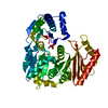
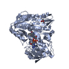
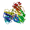
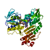
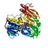
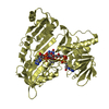
| Title | Crystal Structure of Cytochrome P450cam crystallized in the presence of a tethered substrate analog AdaC3-Etg-Boc | ||||||
 Components Components | Camphor 5-monooxygenase | ||||||
 Keywords Keywords | OXIDOREDUCTASE / Cytochrome P450 / P450cam / camphor / tethered substrate analog / open conformation | ||||||
| Function / homology |  Function and homology information Function and homology informationcamphor 5-monooxygenase / camphor 5-monooxygenase activity / (+)-camphor catabolic process / iron ion binding / heme binding / cytoplasm Similarity search - Function | ||||||
| Biological species |  Pseudomonas putida (bacteria) Pseudomonas putida (bacteria) | ||||||
| Method |  X-RAY DIFFRACTION / SYNCHROTRON / MOLECULAR REPLACEMENT / molecular replacement / Resolution: 2.1 Å X-RAY DIFFRACTION / SYNCHROTRON / MOLECULAR REPLACEMENT / molecular replacement / Resolution: 2.1 Å | ||||||
 Authors Authors | Lee, Y.-T. / Wilson, R.F. / Glazer, E.C. / Goodin, D.B. | ||||||
 Citation Citation | Journal: To be Published Title: Crystal Structure of Cytochrome P450cam crystallized in the presence of a tethered substrate analog AdaC3-Etg-Boc Authors: Lee, Y.-T. / Glazer, E.C. / Wilson, R.F. / Goodin, D.B. | ||||||
| History |
|
- Structure visualization
| Structure viewer | Molecule: MolmilJmol/JSmol |
|---|
- Downloads & links
-Download
| PDBx/mmCIF format | 3p6w.cif.gz | 95.9 KB | Display | PDBx/mmCIF format |
|---|---|---|---|---|
| PDB format | pdb3p6w.ent.gz | 72.2 KB | Display | PDB format |
| PDBx/mmJSON format | 3p6w.json.gz | Tree view | PDBx/mmJSON format | |
| Others |  Other downloads Other downloads |
-Validation report
| Arichive directory | https://data.pdbj.org/pub/pdb/validation_reports/p6/3p6wftp://data.pdbj.org/pub/pdb/validation_reports/p6/3p6w | HTTPS FTP |
|---|
-Related structure data
| Related structure data | |
|---|---|
| Similar structure data |


-Links
 PDBj
PDBj
- Assembly
| Deposited unit | 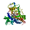
| ||||||||
|---|---|---|---|---|---|---|---|---|---|
| 1 |
| ||||||||
| Unit cell |
|
-Components
| #1: Protein | Mass: 46556.816 Da / Num. of mol.: 1 / Mutation: C334A Source method: isolated from a genetically manipulated source Source: (gene. exp.) Pseudomonas putida (bacteria) / Gene: camC, cyp101 / Plasmid: pET15b / Production host: |
|---|---|
| #2: Chemical | ChemComp-HEM /   Mass: 616.487 Da / Num. of mol.: 1 / Source method: obtained synthetically / Formula: C34H32FeN4O4 Mass: 616.487 Da / Num. of mol.: 1 / Source method: obtained synthetically / Formula: C34H32FeN4O4 |
| #3: Water | ChemComp-HOH /  Mass: 18.015 Da / Num. of mol.: 111 / Source method: isolated from a natural source / Formula: H2O Mass: 18.015 Da / Num. of mol.: 111 / Source method: isolated from a natural source / Formula: H2O |
-Experimental details
-Experiment
| Experiment | Method: X-RAY DIFFRACTION / Number of used crystals: 1 |
|---|
- Sample preparation
| Crystal | Density Matthews: 2.47 Å3/Da / Density % sol: 50.15 % |
|---|---|
| Crystal grow | Temperature: 279 K / Method: sitting-drop vapor diffusion / pH: 6.5 Details: 12-22% PEG 8000, 0.1M Sodium cacodylate, pH 6.5, 0.1-0.2M KCl, sitting-drop vapor diffusion, temperature 279K |
-Data collection
| Diffraction | Mean temperature: 100 K | ||||||||||||||||||||||||||||||||||||||||||||||||||||||||||||||||||||||||||||||||||||||||
|---|---|---|---|---|---|---|---|---|---|---|---|---|---|---|---|---|---|---|---|---|---|---|---|---|---|---|---|---|---|---|---|---|---|---|---|---|---|---|---|---|---|---|---|---|---|---|---|---|---|---|---|---|---|---|---|---|---|---|---|---|---|---|---|---|---|---|---|---|---|---|---|---|---|---|---|---|---|---|---|---|---|---|---|---|---|---|---|---|---|
| Diffraction source | Source: SYNCHROTRON / Site: SSRL  / Beamline: BL7-1 / Wavelength: 0.97839 Å / Beamline: BL7-1 / Wavelength: 0.97839 Å | ||||||||||||||||||||||||||||||||||||||||||||||||||||||||||||||||||||||||||||||||||||||||
| Detector | Type: ADSC QUANTUM 315r / Detector: CCD / Date: Jul 6, 2007 / Details: RH COATED FLAT MIRROR | ||||||||||||||||||||||||||||||||||||||||||||||||||||||||||||||||||||||||||||||||||||||||
| Radiation | Monochromator: SIDE SCATTERING I-BEAM BENT SINGLE CRYSTAL / Protocol: SINGLE WAVELENGTH / Monochromatic (M) / Laue (L): M / Scattering type: x-ray | ||||||||||||||||||||||||||||||||||||||||||||||||||||||||||||||||||||||||||||||||||||||||
| Radiation wavelength | Wavelength: 0.97839 Å / Relative weight: 1 | ||||||||||||||||||||||||||||||||||||||||||||||||||||||||||||||||||||||||||||||||||||||||
| Reflection | Resolution: 2.1→92.946 Å / Num. all: 27619 / Num. obs: 25134 / % possible obs: 91.8 % / Redundancy: 4.5 % / Biso Wilson estimate: 33.9 Å2 / Rsym value: 0.085 / Net I/σ(I): 9.8 | ||||||||||||||||||||||||||||||||||||||||||||||||||||||||||||||||||||||||||||||||||||||||
| Reflection shell |
|
-Phasing
| Phasing | Method: molecular replacement | |||||||||
|---|---|---|---|---|---|---|---|---|---|---|
| Phasing MR |
|
- Processing
| Software |
| |||||||||||||||||||||||||||||||||||||||||||||||||||||||||||||||||
|---|---|---|---|---|---|---|---|---|---|---|---|---|---|---|---|---|---|---|---|---|---|---|---|---|---|---|---|---|---|---|---|---|---|---|---|---|---|---|---|---|---|---|---|---|---|---|---|---|---|---|---|---|---|---|---|---|---|---|---|---|---|---|---|---|---|---|
| Refinement | Method to determine structure: MOLECULAR REPLACEMENT / Resolution: 2.1→10 Å / Cor.coef. Fo:Fc: 0.949 / Cor.coef. Fo:Fc free: 0.91 / WRfactor Rfree: 0.2562 / WRfactor Rwork: 0.2011 / Occupancy max: 1 / Occupancy min: 0.5 / FOM work R set: 0.8182 / SU B: 5.355 / SU ML: 0.143 / SU R Cruickshank DPI: 0.2602 / SU Rfree: 0.2187 / Cross valid method: THROUGHOUT / σ(F): 0 / ESU R Free: 0.219 / Stereochemistry target values: MAXIMUM LIKELIHOOD Details: HYDROGENS HAVE BEEN ADDED IN THE RIDING POSITIONS U VALUES : REFINED INDIVIDUALLY
| |||||||||||||||||||||||||||||||||||||||||||||||||||||||||||||||||
| Solvent computation | Ion probe radii: 0.8 Å / Shrinkage radii: 0.8 Å / VDW probe radii: 1.2 Å / Solvent model: MASK | |||||||||||||||||||||||||||||||||||||||||||||||||||||||||||||||||
| Displacement parameters | Biso max: 62.02 Å2 / Biso mean: 34.3977 Å2 / Biso min: 18.9 Å2
| |||||||||||||||||||||||||||||||||||||||||||||||||||||||||||||||||
| Refinement step | Cycle: LAST / Resolution: 2.1→10 Å
| |||||||||||||||||||||||||||||||||||||||||||||||||||||||||||||||||
| Refine LS restraints |
| |||||||||||||||||||||||||||||||||||||||||||||||||||||||||||||||||
| LS refinement shell | Resolution: 2.1→2.152 Å / Total num. of bins used: 20
|
