 Movie
Movie Controller


+ Open data

- Basic information
| Entry | Database: PDB / ID: 1f3p | ||||||
|---|---|---|---|---|---|---|---|
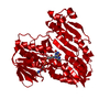
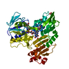
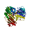
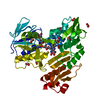
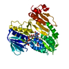
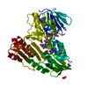
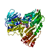
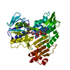
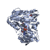
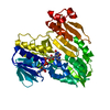
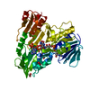
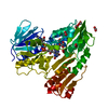
| Title | FERREDOXIN REDUCTASE (BPHA4)-NADH COMPLEX | ||||||
 Components Components | FERREDOXIN REDUCTASE | ||||||
 Keywords Keywords | OXIDOREDUCTASE / Rossmann fold | ||||||
| Function / homology |  Function and homology information Function and homology informationoxidoreductase activity, acting on NAD(P)H / nucleotide binding / cytoplasm Similarity search - Function | ||||||
| Biological species |  Pseudomonas sp. (bacteria) Pseudomonas sp. (bacteria) | ||||||
| Method |  X-RAY DIFFRACTION / SYNCHROTRON / Resolution: 2.4 Å X-RAY DIFFRACTION / SYNCHROTRON / Resolution: 2.4 Å | ||||||
 Authors Authors | Senda, T. / Yamada, T. / Sakurai, N. / Kubota, M. / Nishizaki, T. / Masai, E. / Fukuda, M. / Mitsuidagger, Y. | ||||||
 Citation Citation | Journal: J.Mol.Biol. / Year: 2000 Title: Crystal structure of NADH-dependent ferredoxin reductase component in biphenyl dioxygenase. Authors: Senda, T. / Yamada, T. / Sakurai, N. / Kubota, M. / Nishizaki, T. / Masai, E. / Fukuda, M. / Mitsuidagger, Y. #1: Journal: J.Bacteriol. / Year: 1994Title: Identification of the bphA4 gene encoding ferredoxin reductase involved in biphenyl and polychlorinated biphenyl degradation in Pseudomonas sp. strain KKS102. Authors: Kikuchi, Y. / Nagata, Y. / Hinata, M. / Kimbara, K. / Fukuda, M. / Yano, Y. / Takagi, M. | ||||||
| History |
|
- Structure visualization
| Structure viewer | Molecule: MolmilJmol/JSmol |
|---|
- Downloads & links
-Download
| PDBx/mmCIF format | 1f3p.cif.gz | 91.3 KB | Display | PDBx/mmCIF format |
|---|---|---|---|---|
| PDB format | pdb1f3p.ent.gz | 69 KB | Display | PDB format |
| PDBx/mmJSON format | 1f3p.json.gz | Tree view | PDBx/mmJSON format | |
| Others |  Other downloads Other downloads |
-Validation report
| Arichive directory | https://data.pdbj.org/pub/pdb/validation_reports/f3/1f3pftp://data.pdbj.org/pub/pdb/validation_reports/f3/1f3p | HTTPS FTP |
|---|
-Related structure data

-Links
 PDBj
PDBj


- Assembly
| Deposited unit | 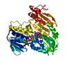
| ||||||||
|---|---|---|---|---|---|---|---|---|---|
| 1 |
| ||||||||
| Unit cell |
| ||||||||
| Details | The biological assembly seems to be a dimer. However, the dimer form can not be indentified in the crystal. |
-Components
| #1: Protein | Mass: 43221.184 Da / Num. of mol.: 1 Source method: isolated from a genetically manipulated source Source: (gene. exp.) Pseudomonas sp. (bacteria) / Strain: KKS102 / Plasmid: PKH204 / Production host: References: UniProt: Q52437, Oxidoreductases; Acting on paired donors, with incorporation or reduction of molecular oxygen |
|---|---|
| #2: Chemical | ChemComp-FAD /   Mass: 785.550 Da / Num. of mol.: 1 / Source method: obtained synthetically / Formula: C27H33N9O15P2 / Comment: FAD*YM Mass: 785.550 Da / Num. of mol.: 1 / Source method: obtained synthetically / Formula: C27H33N9O15P2 / Comment: FAD*YM |
| #3: Chemical | ChemComp-NAD /   Mass: 663.425 Da / Num. of mol.: 1 / Source method: obtained synthetically / Formula: C21H27N7O14P2 / Comment: NAD*YM Mass: 663.425 Da / Num. of mol.: 1 / Source method: obtained synthetically / Formula: C21H27N7O14P2 / Comment: NAD*YM |
| #4: Water | ChemComp-HOH /  Mass: 18.015 Da / Num. of mol.: 81 / Source method: isolated from a natural source / Formula: H2O Mass: 18.015 Da / Num. of mol.: 81 / Source method: isolated from a natural source / Formula: H2O |
-Experimental details
-Experiment
| Experiment | Method: X-RAY DIFFRACTION / Number of used crystals: 1 |
|---|
- Sample preparation
| Crystal | Density Matthews: 2.79 Å3/Da / Density % sol: 55.91 % | ||||||||||||||||||||
|---|---|---|---|---|---|---|---|---|---|---|---|---|---|---|---|---|---|---|---|---|---|
| Crystal grow | Temperature: 293 K / Method: vapor diffusion, hanging drop / pH: 4.6 Details: 2.0M Sodium formate, 100mM Na acetate buffer, pH 4.6, VAPOR DIFFUSION, HANGING DROP, temperature 293K | ||||||||||||||||||||
| Crystal grow | *PLUS Temperature: 20 ℃ / pH: 5.4 / Details: Yamada, T., (2000) Protein Pept.Lett., 7, 277. | ||||||||||||||||||||
| Components of the solutions | *PLUS
|
-Data collection
| Diffraction | Mean temperature: 100 K |
|---|---|
| Diffraction source | Source: SYNCHROTRON / Site: Photon Factory  / Beamline: BL-18B / Wavelength: 1 / Beamline: BL-18B / Wavelength: 1 |
| Detector | Type: WEISSENBERG / Detector: DIFFRACTOMETER / Date: Dec 11, 1998 |
| Radiation | Protocol: SINGLE WAVELENGTH / Monochromatic (M) / Laue (L): M / Scattering type: x-ray |
| Radiation wavelength | Wavelength: 1 Å / Relative weight: 1 |
| Reflection | Resolution: 2.4→38 Å / Num. all: 133802 / Num. obs: 133802 / % possible obs: 99.6 % / Observed criterion σ(F): 0 / Observed criterion σ(I): 0 / Redundancy: 6.7 % / Biso Wilson estimate: 41.4 Å2 / Rmerge(I) obs: 0.085 / Net I/σ(I): 8.9 |
| Reflection shell | Resolution: 2.4→2.53 Å / Redundancy: 6 % / Rmerge(I) obs: 0.478 / % possible all: 99.2 |
| Reflection | *PLUS Num. obs: 19786 / Num. measured all: 133802 |
- Processing
| Software |
| ||||||||||||||||||||
|---|---|---|---|---|---|---|---|---|---|---|---|---|---|---|---|---|---|---|---|---|---|
| Refinement | Resolution: 2.4→38 Å / σ(F): 1 / σ(I): 1 / Stereochemistry target values: Engh & Huber / Details: Used bulk solvent correction.
| ||||||||||||||||||||
| Refinement step | Cycle: LAST / Resolution: 2.4→38 Å
| ||||||||||||||||||||
| Refine LS restraints |
| ||||||||||||||||||||
| Software | *PLUS Name: X-PLOR / Version: 3.851 / Classification: refinement | ||||||||||||||||||||
| Refinement | *PLUS Highest resolution: 2.4 Å / Lowest resolution: 38 Å / σ(F): 2 / % reflection Rfree: 10 % / Rfactor obs: 0.206 | ||||||||||||||||||||
| Solvent computation | *PLUS | ||||||||||||||||||||
| Displacement parameters | *PLUS | ||||||||||||||||||||
| Refine LS restraints | *PLUS
|
