 Movie
Movie Controller

[English] 日本語
 Yorodumi
- PDB-4h4t: Crystal Structure of Ferredoxin reductase, BphA4 T176R mutant (ox... -
+ Open data

- Basic information
| Entry | Database: PDB / ID: 4h4t | ||||||
|---|---|---|---|---|---|---|---|
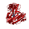
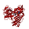
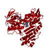
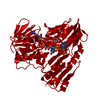
| Title | Crystal Structure of Ferredoxin reductase, BphA4 T176R mutant (oxidized form) | ||||||
 Components Components | Biphenyl dioxygenase ferredoxin reductase subunit | ||||||
 Keywords Keywords | OXIDOREDUCTASE / Flavoprotein | ||||||
| Function / homology |  Function and homology information Function and homology informationdioxygenase activity / oxidoreductase activity, acting on NAD(P)H / nucleotide binding / cytoplasm Similarity search - Function | ||||||
| Biological species |  Acidovorax (bacteria) Acidovorax (bacteria) | ||||||
| Method |  X-RAY DIFFRACTION / SYNCHROTRON / MOLECULAR REPLACEMENT / Resolution: 1.5 Å X-RAY DIFFRACTION / SYNCHROTRON / MOLECULAR REPLACEMENT / Resolution: 1.5 Å | ||||||
 Authors Authors | Nishizawa, A. / Harada, A. / Senda, M. / Tachihara, Y. / Muramatsu, D. / Kishigami, S. / Mori, S. / Sugiyama, K. / Senda, T. / Kimura, S. | ||||||
 Citation Citation | Journal: To be Published Title: Random Mutagenesis with the Project Assessment for Complete Conversion of Co-Factor Specificity of a Ferredoxin Reductase BphA4 Authors: Nishizawa, A. / Harada, A. / Senda, M. / Tachihara, Y. / Muramatsu, D. / Kishigami, S. / Mori, S. / Sugiyama, K. / Senda, T. / Kimura, S. | ||||||
| History |
|
- Structure visualization
| Structure viewer | Molecule: MolmilJmol/JSmol |
|---|
- Downloads & links
-Download
| PDBx/mmCIF format | 4h4t.cif.gz | 101.8 KB | Display | PDBx/mmCIF format |
|---|---|---|---|---|
| PDB format | pdb4h4t.ent.gz | 75.4 KB | Display | PDB format |
| PDBx/mmJSON format | 4h4t.json.gz | Tree view | PDBx/mmJSON format | |
| Others |  Other downloads Other downloads |
-Validation report
| Arichive directory | https://data.pdbj.org/pub/pdb/validation_reports/h4/4h4tftp://data.pdbj.org/pub/pdb/validation_reports/h4/4h4t | HTTPS FTP |
|---|
-Related structure data
| Related structure data | 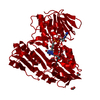 4h4pC  4h4qC 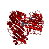 4h4rC 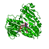 4h4sC  4h4uC 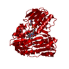 4h4vC  4h4wC  4h4xC  4h4yC  4h4zC  4h50C  2gqwS C: citing same article ( S: Starting model for refinement |
|---|---|
| Similar structure data |


-Links
 PDBj
PDBj


- Assembly
| Deposited unit | 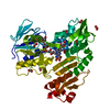
| ||||||||
|---|---|---|---|---|---|---|---|---|---|
| 1 | 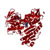
| ||||||||
| Unit cell |
|
-Components
| #1: Protein | Mass: 43277.273 Da / Num. of mol.: 1 / Mutation: T176R Source method: isolated from a genetically manipulated source Source: (gene. exp.) Acidovorax (bacteria) / Strain: KKS102 / Gene: bphA4 / Production host: | ||
|---|---|---|---|
| #2: Chemical | ChemComp-FAD /   Mass: 785.550 Da / Num. of mol.: 1 / Source method: obtained synthetically / Formula: C27H33N9O15P2 / Comment: FAD*YM Mass: 785.550 Da / Num. of mol.: 1 / Source method: obtained synthetically / Formula: C27H33N9O15P2 / Comment: FAD*YM | ||
| #3: Chemical | ChemComp-GOL /   Mass: 92.094 Da / Num. of mol.: 1 / Source method: obtained synthetically / Formula: C3H8O3 Mass: 92.094 Da / Num. of mol.: 1 / Source method: obtained synthetically / Formula: C3H8O3 | ||
| #4: Chemical | ChemComp-FMT /   Mass: 46.025 Da / Num. of mol.: 12 / Source method: obtained synthetically / Formula: CH2O2 Mass: 46.025 Da / Num. of mol.: 12 / Source method: obtained synthetically / Formula: CH2O2#5: Water | ChemComp-HOH / |  Mass: 18.015 Da / Num. of mol.: 397 / Source method: isolated from a natural source / Formula: H2O Mass: 18.015 Da / Num. of mol.: 397 / Source method: isolated from a natural source / Formula: H2O |
-Experimental details
-Experiment
| Experiment | Method: X-RAY DIFFRACTION / Number of used crystals: 1 |
|---|
- Sample preparation
| Crystal | Density Matthews: 2.75 Å3/Da / Density % sol: 55.24 % |
|---|---|
| Crystal grow | Temperature: 293 K / Method: vapor diffusion, sitting drop / pH: 5.2 Details: 2.5M Sodium formate, 0.1M acetate buffer, pH 5.2, VAPOR DIFFUSION, SITTING DROP, temperature 293K |
-Data collection
| Diffraction | Mean temperature: 95 K |
|---|---|
| Diffraction source | Source: SYNCHROTRON / Site: Photon Factory  / Beamline: AR-NE3A / Wavelength: 0.9796 Å / Beamline: AR-NE3A / Wavelength: 0.9796 Å |
| Detector | Type: ADSC QUANTUM 270 / Detector: CCD / Date: Oct 10, 2010 |
| Radiation | Monochromator: Si 111 / Protocol: SINGLE WAVELENGTH / Monochromatic (M) / Laue (L): M / Scattering type: x-ray |
| Radiation wavelength | Wavelength: 0.9796 Å / Relative weight: 1 |
| Reflection | Resolution: 1.5→85.16 Å / Num. all: 77482 / Num. obs: 77482 / % possible obs: 99 % / Observed criterion σ(F): 0 / Observed criterion σ(I): 0 / Biso Wilson estimate: 13.6 Å2 |
| Reflection shell | Resolution: 1.5→1.58 Å / % possible all: 98.6 |
- Processing
| Software |
| |||||||||||||||||||||||||||||||||||||||||||||||||||||||||||||||||||||||||||||||||||||||||||||||||||||||||||||||||||||||||||||||||||||||||||||||||||||||||||||||||||||||||||||||||||||||||||||||||||||||||||
|---|---|---|---|---|---|---|---|---|---|---|---|---|---|---|---|---|---|---|---|---|---|---|---|---|---|---|---|---|---|---|---|---|---|---|---|---|---|---|---|---|---|---|---|---|---|---|---|---|---|---|---|---|---|---|---|---|---|---|---|---|---|---|---|---|---|---|---|---|---|---|---|---|---|---|---|---|---|---|---|---|---|---|---|---|---|---|---|---|---|---|---|---|---|---|---|---|---|---|---|---|---|---|---|---|---|---|---|---|---|---|---|---|---|---|---|---|---|---|---|---|---|---|---|---|---|---|---|---|---|---|---|---|---|---|---|---|---|---|---|---|---|---|---|---|---|---|---|---|---|---|---|---|---|---|---|---|---|---|---|---|---|---|---|---|---|---|---|---|---|---|---|---|---|---|---|---|---|---|---|---|---|---|---|---|---|---|---|---|---|---|---|---|---|---|---|---|---|---|---|---|---|---|---|---|
| Refinement | Method to determine structure: MOLECULAR REPLACEMENT Starting model: 2GQW Resolution: 1.5→76.177 Å / Occupancy max: 1 / Occupancy min: 0.26 / FOM work R set: 0.8703 / SU ML: 0.14 / σ(F): 1.99 / Phase error: 19.63 / Stereochemistry target values: ML
| |||||||||||||||||||||||||||||||||||||||||||||||||||||||||||||||||||||||||||||||||||||||||||||||||||||||||||||||||||||||||||||||||||||||||||||||||||||||||||||||||||||||||||||||||||||||||||||||||||||||||||
| Solvent computation | Shrinkage radii: 0.9 Å / VDW probe radii: 1.11 Å / Solvent model: FLAT BULK SOLVENT MODEL | |||||||||||||||||||||||||||||||||||||||||||||||||||||||||||||||||||||||||||||||||||||||||||||||||||||||||||||||||||||||||||||||||||||||||||||||||||||||||||||||||||||||||||||||||||||||||||||||||||||||||||
| Displacement parameters | Biso max: 56.54 Å2 / Biso mean: 17.3861 Å2 / Biso min: 6.04 Å2 | |||||||||||||||||||||||||||||||||||||||||||||||||||||||||||||||||||||||||||||||||||||||||||||||||||||||||||||||||||||||||||||||||||||||||||||||||||||||||||||||||||||||||||||||||||||||||||||||||||||||||||
| Refinement step | Cycle: LAST / Resolution: 1.5→76.177 Å
| |||||||||||||||||||||||||||||||||||||||||||||||||||||||||||||||||||||||||||||||||||||||||||||||||||||||||||||||||||||||||||||||||||||||||||||||||||||||||||||||||||||||||||||||||||||||||||||||||||||||||||
| Refine LS restraints |
| |||||||||||||||||||||||||||||||||||||||||||||||||||||||||||||||||||||||||||||||||||||||||||||||||||||||||||||||||||||||||||||||||||||||||||||||||||||||||||||||||||||||||||||||||||||||||||||||||||||||||||
| LS refinement shell | Refine-ID: X-RAY DIFFRACTION / Total num. of bins used: 28
|
