 Movie
Movie Controller

[English] 日本語
 Yorodumi
- PDB-7btj: Crystal structure of Pennisetum glaucum monodehydroascorbate redu... -
+ Open data

- Basic information
| Entry | Database: PDB / ID: 7btj | |||||||||
|---|---|---|---|---|---|---|---|---|---|---|
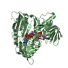
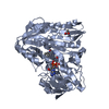
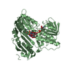
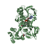
| Title | Crystal structure of Pennisetum glaucum monodehydroascorbate reductase in complex with FADH2 | |||||||||
 Components Components | Pennisetum glaucum monodehydroascorbate reductase | |||||||||
 Keywords Keywords | OXIDOREDUCTASE / Nucleotide Binding Oxidoreductase Activity Monodehydroascorbate Reductase (nadh) Activity Flavin Adenine Dinucleotide Binding | |||||||||
| Function / homology | DIHYDROFLAVINE-ADENINE DINUCLEOTIDE / NICOTINAMIDE-ADENINE-DINUCLEOTIDE Function and homology information Function and homology information | |||||||||
| Biological species |  Cenchrus americanus (pearl millet) Cenchrus americanus (pearl millet) | |||||||||
| Method |  X-RAY DIFFRACTION / MOLECULAR REPLACEMENT / Resolution: 2.373 Å X-RAY DIFFRACTION / MOLECULAR REPLACEMENT / Resolution: 2.373 Å | |||||||||
 Authors Authors | Sonkar, K.S. / Achary, M.M. / Reddy, M.K. / Arulandu, A. | |||||||||
| Funding support |  India, 1items India, 1items
| |||||||||
 Citation Citation | Journal: To Be Published Title: Crystal structure of Pennisetum glaucum monodehydroascorbate reductase Authors: Sonkar, K.S. / Arulandu, A. / Achary, M.M. / Reddy, M.K. | |||||||||
| History |
|
- Structure visualization
| Structure viewer | Molecule: MolmilJmol/JSmol |
|---|
- Downloads & links
-Download
| PDBx/mmCIF format | 7btj.cif.gz | 347.5 KB | Display | PDBx/mmCIF format |
|---|---|---|---|---|
| PDB format | pdb7btj.ent.gz | 279.4 KB | Display | PDB format |
| PDBx/mmJSON format | 7btj.json.gz | Tree view | PDBx/mmJSON format | |
| Others |  Other downloads Other downloads |
-Validation report
| Arichive directory | https://data.pdbj.org/pub/pdb/validation_reports/bt/7btjftp://data.pdbj.org/pub/pdb/validation_reports/bt/7btj | HTTPS FTP |
|---|
-Related structure data
| Related structure data | 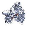 7buzC 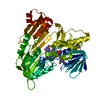 5jciS S: Starting model for refinement C: citing same article ( |
|---|---|
| Similar structure data |


-Links
 PDBj
PDBj- Assembly
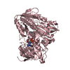
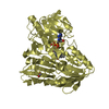
| Deposited unit | 
| ||||||||
|---|---|---|---|---|---|---|---|---|---|
| 1 | 
| ||||||||
| 2 | 
| ||||||||
| 3 | 
| ||||||||
| 4 | 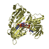
| ||||||||
| Unit cell |
|
-Components
| #1: Protein | Mass: 46843.133 Da / Num. of mol.: 4 Source method: isolated from a genetically manipulated source Source: (gene. exp.) Cenchrus americanus (pearl millet) / Gene: MDHAR / Plasmid: pETM30 / Production host:  #2: Chemical | ChemComp-NAD /   Mass: 663.425 Da / Num. of mol.: 4 / Source method: obtained synthetically / Formula: C21H27N7O14P2 / Comment: NAD*YM Mass: 663.425 Da / Num. of mol.: 4 / Source method: obtained synthetically / Formula: C21H27N7O14P2 / Comment: NAD*YM#3: Chemical | ChemComp-FDA /   Mass: 787.566 Da / Num. of mol.: 4 / Source method: obtained synthetically / Formula: C27H35N9O15P2 / Feature type: SUBJECT OF INVESTIGATION Mass: 787.566 Da / Num. of mol.: 4 / Source method: obtained synthetically / Formula: C27H35N9O15P2 / Feature type: SUBJECT OF INVESTIGATION#4: Water | ChemComp-HOH / |  Mass: 18.015 Da / Num. of mol.: 553 / Source method: isolated from a natural source / Formula: H2O Mass: 18.015 Da / Num. of mol.: 553 / Source method: isolated from a natural source / Formula: H2OHas ligand of interest | Y | |
|---|
-Experimental details
-Experiment
| Experiment | Method: X-RAY DIFFRACTION / Number of used crystals: 1 |
|---|
- Sample preparation
| Crystal | Density Matthews: 2.19 Å3/Da / Density % sol: 45 % |
|---|---|
| Crystal grow | Temperature: 293.15 K / Method: vapor diffusion, sitting drop / pH: 6.5 Details: 0.2 M Sodium acetate trihydrate 0.1 M Sodium cacodylate trihydrate pH 6.5 30% w/v Polyethylene glycol 8,000 PH range: 6.4 - 6.8 |
-Data collection
| Diffraction | Mean temperature: 100 K / Serial crystal experiment: N |
|---|---|
| Diffraction source | Source: ROTATING ANODE / Type: RIGAKU MICROMAX-007 / Wavelength: 1.5418 Å |
| Detector | Type: MAR scanner 345 mm plate / Detector: IMAGE PLATE / Date: Jan 18, 2019 |
| Radiation | Protocol: SINGLE WAVELENGTH / Monochromatic (M) / Laue (L): M / Scattering type: x-ray |
| Radiation wavelength | Wavelength: 1.5418 Å / Relative weight: 1 |
| Reflection | Resolution: 2.37→61.45 Å / Num. obs: 64802 / % possible obs: 96.6 % / Redundancy: 4.9 % / CC1/2: 0.997 / Net I/σ(I): 2.21 |
| Reflection shell | Resolution: 2.373→2.413 Å / Num. unique obs: 15670 / CC1/2: 0.997 |
- Processing
| Software |
| ||||||||||||||||||||||||||||||||||||||||||||||||||||||||||||||||||||||||||||||||||||||||||||||||||||||||||||
|---|---|---|---|---|---|---|---|---|---|---|---|---|---|---|---|---|---|---|---|---|---|---|---|---|---|---|---|---|---|---|---|---|---|---|---|---|---|---|---|---|---|---|---|---|---|---|---|---|---|---|---|---|---|---|---|---|---|---|---|---|---|---|---|---|---|---|---|---|---|---|---|---|---|---|---|---|---|---|---|---|---|---|---|---|---|---|---|---|---|---|---|---|---|---|---|---|---|---|---|---|---|---|---|---|---|---|---|---|---|
| Refinement | Method to determine structure: MOLECULAR REPLACEMENT Starting model: 5JCI Resolution: 2.373→61.45 Å / Cor.coef. Fo:Fc: 0.936 / Cor.coef. Fo:Fc free: 0.899 / SU R Cruickshank DPI: 0.532 / Cross valid method: THROUGHOUT / σ(F): 0 / SU R Blow DPI: 0.488 / SU Rfree Blow DPI: 0.261 / SU Rfree Cruickshank DPI: 0.269
| ||||||||||||||||||||||||||||||||||||||||||||||||||||||||||||||||||||||||||||||||||||||||||||||||||||||||||||
| Displacement parameters | Biso max: 92.79 Å2 / Biso mean: 41.01 Å2 / Biso min: 14.42 Å2
| ||||||||||||||||||||||||||||||||||||||||||||||||||||||||||||||||||||||||||||||||||||||||||||||||||||||||||||
| Refine analyze | Luzzati coordinate error obs: 0.28 Å | ||||||||||||||||||||||||||||||||||||||||||||||||||||||||||||||||||||||||||||||||||||||||||||||||||||||||||||
| Refinement step | Cycle: final / Resolution: 2.373→61.45 Å
| ||||||||||||||||||||||||||||||||||||||||||||||||||||||||||||||||||||||||||||||||||||||||||||||||||||||||||||
| Refine LS restraints |
| ||||||||||||||||||||||||||||||||||||||||||||||||||||||||||||||||||||||||||||||||||||||||||||||||||||||||||||
| LS refinement shell | Resolution: 2.39→2.39 Å / Rfactor Rfree error: 0
|
