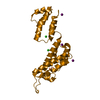
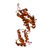
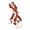
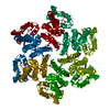
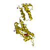
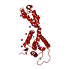
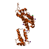
+Open data
-Basic information
Entry | Database: PDB / ID: 6mqo | ||||||
---|---|---|---|---|---|---|---|
Title | Structure of HIV-1 CA G208R | ||||||
Components | Capsid protein | ||||||
Keywords | VIRAL PROTEIN / Capsid | ||||||
Function / homology | Function and homology information viral budding via host ESCRT complex / viral process / ISG15 antiviral mechanism / host multivesicular body / viral nucleocapsid / host cell cytoplasm / host cell nucleus / host cell plasma membrane / structural molecule activity / virion membrane ...viral budding via host ESCRT complex / viral process / ISG15 antiviral mechanism / host multivesicular body / viral nucleocapsid / host cell cytoplasm / host cell nucleus / host cell plasma membrane / structural molecule activity / virion membrane / RNA binding / zinc ion binding / ATP binding / membrane / cytoplasm Similarity search - Function | ||||||
Biological species | Human immunodeficiency virus 1 | ||||||
Method | X-RAY DIFFRACTION / SYNCHROTRON / MOLECULAR REPLACEMENT / Resolution: 3.2 Å | ||||||
Authors | Smaga, S.S. / Xiong, Y. | ||||||
Funding support | United States, 1items
| ||||||
Citation | Journal: Structure / Year: 2019 Title: MxB Restricts HIV-1 by Targeting the Tri-hexamer Interface of the Viral Capsid. Authors: Smaga, S.S. / Xu, C. / Summers, B.J. / Digianantonio, K.M. / Perilla, J.R. / Xiong, Y. | ||||||
History |
|
-Structure visualization
Structure viewer | Molecule: MolmilJmol/JSmol |
---|
-Downloads & links
-Download
PDBx/mmCIF format | 6mqo.cif.gz | 102.3 KB | Display | PDBx/mmCIF format |
---|---|---|---|---|
PDB format | pdb6mqo.ent.gz | 77.2 KB | Display | PDB format |
PDBx/mmJSON format | 6mqo.json.gz | Tree view | PDBx/mmJSON format | |
Others | Other downloads |
-Validation report
Arichive directory | https://data.pdbj.org/pub/pdb/validation_reports/mq/6mqo ftp://data.pdbj.org/pub/pdb/validation_reports/mq/6mqo | HTTPS FTP |
---|
-Related structure data
Related structure data | 6mqaC 6mqpC 4xfxS S: Starting model for refinement C: citing same article (ref.) |
---|---|
Similar structure data |
-Links
-Assembly
Deposited unit |
| ||||||||
---|---|---|---|---|---|---|---|---|---|
1 |
| x 6||||||||
Unit cell |
|
-Components
#1: Protein | Mass: 25861.766 Da / Num. of mol.: 1 / Fragment: UNP residues 133-363 / Mutation: G208R Source method: isolated from a genetically manipulated source Source: (gene. exp.) Human immunodeficiency virus 1 / Gene: gag / Plasmid: pET11 / Production host: Escherichia coli BL21(DE3) (bacteria) / References: UniProt: B6DRA0, UniProt: P12493*PLUS | ||
---|---|---|---|
#2: Chemical | #3: Water | ChemComp-HOH / | |
-Experimental details
-Experiment
Experiment | Method: X-RAY DIFFRACTION / Number of used crystals: 1 |
---|
-Sample preparation
Crystal | Density Matthews: 2.7 Å3/Da / Density % sol: 54.39 % |
---|---|
Crystal grow | Temperature: 293 K / Method: microbatch / pH: 6.5 Details: 15% PEG3350, 0.1 M sodium iodide, Bis-Tris propane, pH 6.5 |
-Data collection
Diffraction | Mean temperature: 100 K / Serial crystal experiment: N |
---|---|
Diffraction source | Source: SYNCHROTRON / Site: APS / Beamline: 24-ID-C / Wavelength: 0.9792 Å |
Detector | Type: DECTRIS PILATUS 6M-F / Detector: PIXEL / Date: Aug 17, 2016 |
Radiation | Monochromator: Cryo-cooled double crystal Si(111) / Protocol: SINGLE WAVELENGTH / Monochromatic (M) / Laue (L): M / Scattering type: x-ray |
Radiation wavelength | Wavelength: 0.9792 Å / Relative weight: 1 |
Reflection | Resolution: 3→50 Å / Num. obs: 4626 / % possible obs: 97.4 % / Redundancy: 2.65 % / Rmerge(I) obs: 0.095 / Net I/σ(I): 9.31 |
Reflection shell | Resolution: 3→3.18 Å / Mean I/σ(I) obs: 1.67 / Num. unique obs: 1671 / % possible all: 94.7 |
-Processing
Software |
| ||||||||||||||||||||||||||||||||||||||||||||||||||||||||||||||||||||||||||||||||||||||||||||||||||||||||||||||||||||||||||||||||||||||||||||||||||||||||||||||||||||||||||||||||||||||
---|---|---|---|---|---|---|---|---|---|---|---|---|---|---|---|---|---|---|---|---|---|---|---|---|---|---|---|---|---|---|---|---|---|---|---|---|---|---|---|---|---|---|---|---|---|---|---|---|---|---|---|---|---|---|---|---|---|---|---|---|---|---|---|---|---|---|---|---|---|---|---|---|---|---|---|---|---|---|---|---|---|---|---|---|---|---|---|---|---|---|---|---|---|---|---|---|---|---|---|---|---|---|---|---|---|---|---|---|---|---|---|---|---|---|---|---|---|---|---|---|---|---|---|---|---|---|---|---|---|---|---|---|---|---|---|---|---|---|---|---|---|---|---|---|---|---|---|---|---|---|---|---|---|---|---|---|---|---|---|---|---|---|---|---|---|---|---|---|---|---|---|---|---|---|---|---|---|---|---|---|---|---|---|
Refinement | Method to determine structure: MOLECULAR REPLACEMENT Starting model: PDB entry 4XFX Resolution: 3.2→20 Å / Cor.coef. Fo:Fc: 0.941 / Cor.coef. Fo:Fc free: 0.86 / SU B: 51.366 / SU ML: 0.4 / Cross valid method: THROUGHOUT / ESU R Free: 0.553 / Stereochemistry target values: MAXIMUM LIKELIHOOD / Details: HYDROGENS HAVE BEEN ADDED IN THE RIDING POSITIONS
| ||||||||||||||||||||||||||||||||||||||||||||||||||||||||||||||||||||||||||||||||||||||||||||||||||||||||||||||||||||||||||||||||||||||||||||||||||||||||||||||||||||||||||||||||||||||
Solvent computation | Ion probe radii: 0.8 Å / Shrinkage radii: 0.8 Å / VDW probe radii: 1.2 Å / Solvent model: MASK | ||||||||||||||||||||||||||||||||||||||||||||||||||||||||||||||||||||||||||||||||||||||||||||||||||||||||||||||||||||||||||||||||||||||||||||||||||||||||||||||||||||||||||||||||||||||
Displacement parameters | Biso mean: 87.361 Å2
| ||||||||||||||||||||||||||||||||||||||||||||||||||||||||||||||||||||||||||||||||||||||||||||||||||||||||||||||||||||||||||||||||||||||||||||||||||||||||||||||||||||||||||||||||||||||
Refinement step | Cycle: 1 / Resolution: 3.2→20 Å
| ||||||||||||||||||||||||||||||||||||||||||||||||||||||||||||||||||||||||||||||||||||||||||||||||||||||||||||||||||||||||||||||||||||||||||||||||||||||||||||||||||||||||||||||||||||||
Refine LS restraints |
|