 Movie
Movie Controller

[English] 日本語
 Yorodumi
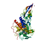
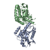
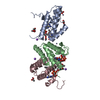
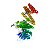
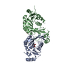
- PDB-6mkh: Crystal structure of pencillin binding protein 4 (PBP4) from Ente... -
+ Open data

- Basic information
| Entry | Database: PDB / ID: 6mkh | ||||||
|---|---|---|---|---|---|---|---|
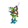
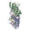
| Title | Crystal structure of pencillin binding protein 4 (PBP4) from Enterococcus faecalis in the imipenem-bound form | ||||||
 Components Components | pencillin binding protein 4 (PBP4) | ||||||
 Keywords Keywords | protein binding/antibiotic / PBP / antibiotic / inhibitor / imipenem / PENICILLIN-BINDING PROTEIN / protein binding-antibiotic complex | ||||||
| Function / homology |  Function and homology information Function and homology informationpeptidoglycan glycosyltransferase / peptidoglycan L,D-transpeptidase activity / glycosyltransferase activity / penicillin binding / cell wall organization / response to antibiotic / plasma membrane Similarity search - Function | ||||||
| Biological species |   Enterococcus faecalis (bacteria) Enterococcus faecalis (bacteria) | ||||||
| Method |  X-RAY DIFFRACTION / SYNCHROTRON / MOLECULAR REPLACEMENT / Resolution: 2.62 Å X-RAY DIFFRACTION / SYNCHROTRON / MOLECULAR REPLACEMENT / Resolution: 2.62 Å | ||||||
 Authors Authors | D'Andrea, E.D. / Moon, T.M. / Peti, W. / Page, R. | ||||||
| Funding support |  United States, 1items United States, 1items
| ||||||
 Citation Citation | Journal: J. Biol. Chem. / Year: 2018 Title: The structures of penicillin-binding protein 4 (PBP4) and PBP5 fromEnterococciprovide structural insights into beta-lactam resistance. Authors: Moon, T.M. / D'Andrea, E.D. / Lee, C.W. / Soares, A. / Jakoncic, J. / Desbonnet, C. / Garcia-Solache, M. / Rice, L.B. / Page, R. / Peti, W. | ||||||
| History |
|
- Structure visualization
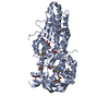
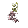
| Structure viewer | Molecule: MolmilJmol/JSmol |
|---|
- Downloads & links
-Download
| PDBx/mmCIF format | 6mkh.cif.gz | 107.1 KB | Display | PDBx/mmCIF format |
|---|---|---|---|---|
| PDB format | pdb6mkh.ent.gz | 76.2 KB | Display | PDB format |
| PDBx/mmJSON format | 6mkh.json.gz | Tree view | PDBx/mmJSON format | |
| Others |  Other downloads Other downloads |
-Validation report
| Arichive directory | https://data.pdbj.org/pub/pdb/validation_reports/mk/6mkhftp://data.pdbj.org/pub/pdb/validation_reports/mk/6mkh | HTTPS FTP |
|---|
-Related structure data
| Related structure data | 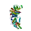 6bsqC 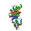 6bsrC 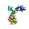 6mkaC 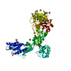 6mkfC 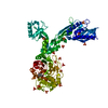 6mkgC 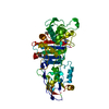 6mkiC 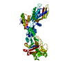 6mkjC C: citing same article ( |
|---|---|
| Similar structure data |

-Links
 PDBj
PDBj

- Assembly
| Deposited unit | 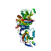
| ||||||||
|---|---|---|---|---|---|---|---|---|---|
| 1 |
| ||||||||
| Unit cell |
|
-Components
| #1: Protein | Mass: 70709.102 Da / Num. of mol.: 1 Source method: isolated from a genetically manipulated source Source: (gene. exp.) Enterococcus faecalis (bacteria) / Gene: pbp4, A6B47_10405, DAI13_12160 / Production host: | ||||
|---|---|---|---|---|---|
| #2: Chemical | ChemComp-IM2 / (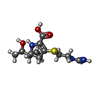  Mass: 301.362 Da / Num. of mol.: 1 / Source method: obtained synthetically / Formula: C12H19N3O4S / Comment: antibiotic*YM Mass: 301.362 Da / Num. of mol.: 1 / Source method: obtained synthetically / Formula: C12H19N3O4S / Comment: antibiotic*YM | ||||
| #3: Chemical |   Mass: 94.971 Da / Num. of mol.: 2 / Source method: obtained synthetically / Formula: PO4 Mass: 94.971 Da / Num. of mol.: 2 / Source method: obtained synthetically / Formula: PO4#4: Water | ChemComp-HOH / |  Mass: 18.015 Da / Num. of mol.: 81 / Source method: isolated from a natural source / Formula: H2O Mass: 18.015 Da / Num. of mol.: 81 / Source method: isolated from a natural source / Formula: H2OHas protein modification | Y | |
-Experimental details
-Experiment
| Experiment | Method: X-RAY DIFFRACTION / Number of used crystals: 1 |
|---|
- Sample preparation
| Crystal | Density Matthews: 2.89 Å3/Da / Density % sol: 57.5 % |
|---|---|
| Crystal grow | Temperature: 294 K / Method: vapor diffusion, hanging drop / pH: 3.25 / Details: 0.04M KH2PO4, 16% PEG 8000, 20% glycerol |
-Data collection
| Diffraction | Mean temperature: 100 K / Serial crystal experiment: N |
|---|---|
| Diffraction source | Source: SYNCHROTRON / Site: SSRL / Beamline: BL12-2 / Wavelength: 0.97946 Å |
| Detector | Type: DECTRIS PILATUS 6M / Detector: PIXEL / Date: Oct 12, 2017 Details: Flat Si Rh coated M0, Kirkpatrick-Baez flat bent Si M1 & M2 |
| Radiation | Monochromator: Liquid nitrogen-cooled double crystal Si(111) Protocol: SINGLE WAVELENGTH / Monochromatic (M) / Laue (L): M / Scattering type: x-ray |
| Radiation wavelength | Wavelength: 0.97946 Å / Relative weight: 1 |
| Reflection | Resolution: 2.62→37.895 Å / Num. obs: 77350 / % possible obs: 96 % / Redundancy: 4.6 % / Biso Wilson estimate: 48.35 Å2 / CC1/2: 0.959 / Rmerge(I) obs: 0.058 / Rpim(I) all: 0.047 / Rrim(I) all: 0.075 / Χ2: 0.72 / Net I/σ(I): 12.5 |
| Reflection shell | Resolution: 2.62→2.74 Å / Redundancy: 4 % / Rmerge(I) obs: 0.211 / Mean I/σ(I) obs: 3.7 / Num. unique obs: 2364 / CC1/2: 0.321 / Rpim(I) all: 0.181 / Rrim(I) all: 0.28 / Χ2: 0.46 / % possible all: 77.3 |
- Processing
| Software |
| ||||||||||||||||||||||||||||||||||||||||||||||||||||||||||||||||||||||
|---|---|---|---|---|---|---|---|---|---|---|---|---|---|---|---|---|---|---|---|---|---|---|---|---|---|---|---|---|---|---|---|---|---|---|---|---|---|---|---|---|---|---|---|---|---|---|---|---|---|---|---|---|---|---|---|---|---|---|---|---|---|---|---|---|---|---|---|---|---|---|---|
| Refinement | Method to determine structure: MOLECULAR REPLACEMENT Starting model: PBP4 apo Resolution: 2.62→37.895 Å / SU ML: 0.25 / Cross valid method: FREE R-VALUE / σ(F): 1.36 / Phase error: 30.38 / Stereochemistry target values: ML
| ||||||||||||||||||||||||||||||||||||||||||||||||||||||||||||||||||||||
| Solvent computation | Shrinkage radii: 0.9 Å / VDW probe radii: 1.11 Å / Solvent model: FLAT BULK SOLVENT MODEL | ||||||||||||||||||||||||||||||||||||||||||||||||||||||||||||||||||||||
| Refinement step | Cycle: LAST / Resolution: 2.62→37.895 Å
| ||||||||||||||||||||||||||||||||||||||||||||||||||||||||||||||||||||||
| Refine LS restraints |
| ||||||||||||||||||||||||||||||||||||||||||||||||||||||||||||||||||||||
| LS refinement shell |
|
