 Movie
Movie Controller

[English] 日本語
 Yorodumi
- PDB-6mic: Crystal Structure of the C-terminal half of the Vibrio cholerae m... -
+ Open data

- Basic information
| Entry | Database: PDB / ID: 6mic | ||||||
|---|---|---|---|---|---|---|---|
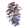
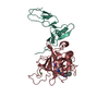
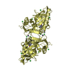
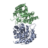
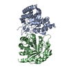
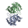
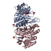
| Title | Crystal Structure of the C-terminal half of the Vibrio cholerae minor pilin TcpB | ||||||
 Components Components | Toxin co-regulated pilus biosynthesis protein B | ||||||
 Keywords Keywords | BIOSYNTHETIC PROTEIN / Effector domain / Minor pilin / Toxin co-regulated pilus / Vibrio cholerae | ||||||
| Function / homology | Toxin co-regulated pilus biosynthesis protein B Function and homology information Function and homology information | ||||||
| Biological species |  Vibrio cholerae serotype O1 (bacteria) Vibrio cholerae serotype O1 (bacteria) | ||||||
| Method |  X-RAY DIFFRACTION / SYNCHROTRON / MOLECULAR REPLACEMENT / Resolution: 1.531 Å X-RAY DIFFRACTION / SYNCHROTRON / MOLECULAR REPLACEMENT / Resolution: 1.531 Å | ||||||
 Authors Authors | Kolappan, S. / Craig, L. | ||||||
| Funding support |  Canada, 1items Canada, 1items
| ||||||
 Citation Citation | Journal: J.Biol.Chem. / Year: 2019 Title: TheVibrio choleraeminor pilin TcpB mediates uptake of the cholera toxin phage CTX phi. Authors: Gutierrez-Rodarte, M. / Kolappan, S. / Burrell, B.A. / Craig, L. | ||||||
| History |
|
- Structure visualization
| Structure viewer | Molecule: MolmilJmol/JSmol |
|---|
- Downloads & links
-Download
| PDBx/mmCIF format | 6mic.cif.gz | 108.9 KB | Display | PDBx/mmCIF format |
|---|---|---|---|---|
| PDB format | pdb6mic.ent.gz | 84.4 KB | Display | PDB format |
| PDBx/mmJSON format | 6mic.json.gz | Tree view | PDBx/mmJSON format | |
| Others |  Other downloads Other downloads |
-Validation report
| Arichive directory | https://data.pdbj.org/pub/pdb/validation_reports/mi/6micftp://data.pdbj.org/pub/pdb/validation_reports/mi/6mic | HTTPS FTP |
|---|
-Related structure data
| Related structure data | 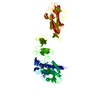 4qs4S S: Starting model for refinement |
|---|---|
| Similar structure data |

-Links
 PDBj
PDBj- Assembly
| Deposited unit | 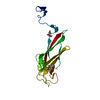
| ||||||||||||
|---|---|---|---|---|---|---|---|---|---|---|---|---|---|
| 1 | 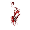
| ||||||||||||
| Unit cell |
| ||||||||||||
| Components on special symmetry positions |
|
-Components
| #1: Protein | Mass: 19642.715 Da / Num. of mol.: 1 Source method: isolated from a genetically manipulated source Source: (gene. exp.) Vibrio cholerae serotype O1 (bacteria) / Strain: ATCC 39541 / Classical Ogawa 395 / O395 / Gene: tcpB, VC0395_A0354 / Production host: | ||||
|---|---|---|---|---|---|
| #2: Chemical | ChemComp-MPD / (  Mass: 118.174 Da / Num. of mol.: 1 / Source method: obtained synthetically / Formula: C6H14O2 / Comment: precipitant*YM Mass: 118.174 Da / Num. of mol.: 1 / Source method: obtained synthetically / Formula: C6H14O2 / Comment: precipitant*YM | ||||
| #3: Chemical |   Mass: 92.094 Da / Num. of mol.: 2 / Source method: obtained synthetically / Formula: C3H8O3 Mass: 92.094 Da / Num. of mol.: 2 / Source method: obtained synthetically / Formula: C3H8O3#4: Water | ChemComp-HOH / |  Mass: 18.015 Da / Num. of mol.: 141 / Source method: isolated from a natural source / Formula: H2O Mass: 18.015 Da / Num. of mol.: 141 / Source method: isolated from a natural source / Formula: H2OHas protein modification | Y | |
-Experimental details
-Experiment
| Experiment | Method: X-RAY DIFFRACTION / Number of used crystals: 1 |
|---|
- Sample preparation
| Crystal | Density Matthews: 2.3 Å3/Da / Density % sol: 47 % |
|---|---|
| Crystal grow | Temperature: 293 K / Method: vapor diffusion, hanging drop / pH: 7.4 / Details: MPD, sodium chloride, sodium acetate / PH range: 4.6 |
-Data collection
| Diffraction | Mean temperature: 100 K |
|---|---|
| Diffraction source | Source: SYNCHROTRON / Site: SSRL  / Beamline: BL9-2 / Wavelength: 1 Å / Beamline: BL9-2 / Wavelength: 1 Å |
| Detector | Type: DECTRIS PILATUS 6M / Detector: PIXEL / Date: Mar 25, 2018 / Details: Rh coated collimating mirror, K-B focusing mirrors |
| Radiation | Protocol: SINGLE WAVELENGTH / Monochromatic (M) / Laue (L): M / Scattering type: x-ray |
| Radiation wavelength | Wavelength: 1 Å / Relative weight: 1 |
| Reflection | Resolution: 1.53→78.8 Å / Num. obs: 26889 / % possible obs: 96.1 % / Redundancy: 3.8 % / Biso Wilson estimate: 12.6 Å2 / CC1/2: 0.999 / Rmerge(I) obs: 0.028 / Rpim(I) all: 0.016 / Rrim(I) all: 0.033 / Χ2: 1.15 / Net I/σ(I): 26.9 |
| Reflection shell | Resolution: 1.53→1.56 Å / Redundancy: 3.8 % / Rmerge(I) obs: 0.091 / Mean I/σ(I) obs: 12.4 / Num. unique obs: 1366 / CC1/2: 0.989 / Rpim(I) all: 0.053 / Rrim(I) all: 0.105 / Χ2: 0.93 / % possible all: 99.4 |
- Processing
| Software |
| |||||||||||||||||||||||||||||||||||||||||||||||||||||||||||||||||||||||||||||
|---|---|---|---|---|---|---|---|---|---|---|---|---|---|---|---|---|---|---|---|---|---|---|---|---|---|---|---|---|---|---|---|---|---|---|---|---|---|---|---|---|---|---|---|---|---|---|---|---|---|---|---|---|---|---|---|---|---|---|---|---|---|---|---|---|---|---|---|---|---|---|---|---|---|---|---|---|---|---|
| Refinement | Method to determine structure: MOLECULAR REPLACEMENT Starting model: 4QS4 Resolution: 1.531→39.382 Å / SU ML: 0.13 / Cross valid method: FREE R-VALUE / σ(F): 1.34 / Phase error: 17.74 / Stereochemistry target values: ML
| |||||||||||||||||||||||||||||||||||||||||||||||||||||||||||||||||||||||||||||
| Solvent computation | Shrinkage radii: 0.9 Å / VDW probe radii: 1.11 Å / Solvent model: FLAT BULK SOLVENT MODEL | |||||||||||||||||||||||||||||||||||||||||||||||||||||||||||||||||||||||||||||
| Refinement step | Cycle: LAST / Resolution: 1.531→39.382 Å
| |||||||||||||||||||||||||||||||||||||||||||||||||||||||||||||||||||||||||||||
| Refine LS restraints |
| |||||||||||||||||||||||||||||||||||||||||||||||||||||||||||||||||||||||||||||
| LS refinement shell |
| |||||||||||||||||||||||||||||||||||||||||||||||||||||||||||||||||||||||||||||
| Refinement TLS params. | Method: refined / Origin x: 25.6022 Å / Origin y: -13.0031 Å / Origin z: 6.9252 Å
| |||||||||||||||||||||||||||||||||||||||||||||||||||||||||||||||||||||||||||||
| Refinement TLS group | Selection details: all |
