 Movie
Movie Controller

[English] 日本語
 Yorodumi
- PDB-6mhy: Structure of connexin-50 intercellular gap junction channel at 3.... -
+ Open data

- Basic information
| Entry | Database: PDB / ID: 6mhy | ||||||
|---|---|---|---|---|---|---|---|
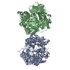
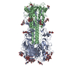
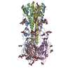
| Title | Structure of connexin-50 intercellular gap junction channel at 3.4 angstrom resolution by cryoEM | ||||||
 Components Components | Gap junction alpha-8 protein, connexin-50 | ||||||
 Keywords Keywords | MEMBRANE PROTEIN / ion channel / gap junction / cell communication / connexin | ||||||
| Function / homology |  Function and homology information Function and homology informationgap junction-mediated intercellular transport / connexin complex / gap junction channel activity / cell-cell signaling / plasma membrane Similarity search - Function | ||||||
| Biological species |  | ||||||
| Method | ELECTRON MICROSCOPY / single particle reconstruction / cryo EM / Resolution: 3.4 Å | ||||||
 Authors Authors | Myers, J.B. / Reichow, S.L. | ||||||
| Funding support |  United States, 1items United States, 1items
| ||||||
 Citation Citation | Journal: Nature / Year: 2018 Title: Structure of native lens connexin 46/50 intercellular channels by cryo-EM. Authors: Janette B Myers / Bassam G Haddad / Susan E O'Neill / Dror S Chorev / Craig C Yoshioka / Carol V Robinson / Daniel M Zuckerman / Steve L Reichow /  Abstract: Gap junctions establish direct pathways for cell-to-cell communication through the assembly of twelve connexin subunits that form intercellular channels connecting neighbouring cells. Co-assembly of ...Gap junctions establish direct pathways for cell-to-cell communication through the assembly of twelve connexin subunits that form intercellular channels connecting neighbouring cells. Co-assembly of different connexin isoforms produces channels with unique properties and enables communication across cell types. Here we used single-particle cryo-electron microscopy to investigate the structural basis of connexin co-assembly in native lens gap junction channels composed of connexin 46 and connexin 50 (Cx46/50). We provide the first comparative analysis to connexin 26 (Cx26), which-together with computational studies-elucidates key energetic features governing gap junction permselectivity. Cx46/50 adopts an open-state conformation that is distinct from the Cx26 crystal structure, yet it appears to be stabilized by a conserved set of hydrophobic anchoring residues. 'Hot spots' of genetic mutations linked to hereditary cataract formation map to the core structural-functional elements identified in Cx46/50, suggesting explanations for many of the disease-causing effects. | ||||||
| History |
|
- Structure visualization
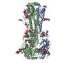
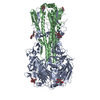
| Movie |
Movie viewer |
|---|---|
| Structure viewer | Molecule: MolmilJmol/JSmol |
- Downloads & links
-Download
| PDBx/mmCIF format | 6mhy.cif.gz | 709 KB | Display | PDBx/mmCIF format |
|---|---|---|---|---|
| PDB format | pdb6mhy.ent.gz | 579.3 KB | Display | PDB format |
| PDBx/mmJSON format | 6mhy.json.gz | Tree view | PDBx/mmJSON format | |
| Others |  Other downloads Other downloads |
-Validation report
| Arichive directory | https://data.pdbj.org/pub/pdb/validation_reports/mh/6mhyftp://data.pdbj.org/pub/pdb/validation_reports/mh/6mhy | HTTPS FTP |
|---|
-Related structure data
| Related structure data | 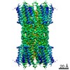 9116MC  6mhqC M: map data used to model this data C: citing same article ( |
|---|---|
| Similar structure data | |
| EM raw data | EMPIAR-10212 (Title: CryoEM reconstruction of native lens connexin-46/50 at 3.4 angstrom resolution Data size: 774.5 Data #1: Unaligned frame stacks - MP38 dataset 01 [micrographs - multiframe] Data #2: Unaligned frame stacks - MP38 dataset 02 [micrographs - multiframe]) |


-Links
 PDBj
PDBj- Assembly
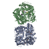
| Deposited unit | 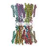
|
|---|---|
| 1 |
|
-Components
| #1: Protein | Mass: 49202.781 Da / Num. of mol.: 12 / Source method: isolated from a natural source / Source: (natural) Plasmid details: C-terminal truncated version isolated from lens core Tissue: Lens / References: UniProt: P55917 Has protein modification | Y | |
|---|
-Experimental details
-Experiment
| Experiment | Method: ELECTRON MICROSCOPY |
|---|---|
| EM experiment | Aggregation state: PARTICLE / 3D reconstruction method: single particle reconstruction |
- Sample preparation
| Component | Name: Connexin-50 gap junction / Type: COMPLEX / Entity ID: all / Source: NATURAL | |||||||||||||||||||||||||
|---|---|---|---|---|---|---|---|---|---|---|---|---|---|---|---|---|---|---|---|---|---|---|---|---|---|---|
| Molecular weight | Value: 0.45 MDa / Experimental value: NO | |||||||||||||||||||||||||
| Source (natural) | Organism: Cellular location: C-terminal truncated version isolated from lens core Organ: Eye / Tissue: Lens | |||||||||||||||||||||||||
| Buffer solution | pH: 7.4 | |||||||||||||||||||||||||
| Buffer component |
| |||||||||||||||||||||||||
| Specimen | Conc.: 2.35 mg/ml / Embedding applied: NO / Shadowing applied: NO / Staining applied: NO / Vitrification applied: YES | |||||||||||||||||||||||||
| Specimen support | Grid material: COPPER / Grid type: Quantifoil, UltrAuFoil, R1.2/1.3 | |||||||||||||||||||||||||
| Vitrification | Instrument: FEI VITROBOT MARK IV / Cryogen name: ETHANE / Humidity: 100 % / Chamber temperature: 298 K |
- Electron microscopy imaging
| Experimental equipment |  Model: Titan Krios / Image courtesy: FEI Company |
|---|---|
| Microscopy | Model: FEI TITAN KRIOS |
| Electron gun | Electron source:  FIELD EMISSION GUN / Accelerating voltage: 300 kV / Illumination mode: FLOOD BEAM FIELD EMISSION GUN / Accelerating voltage: 300 kV / Illumination mode: FLOOD BEAM |
| Electron lens | Mode: BRIGHT FIELD / Nominal magnification: 105000 X / Nominal defocus max: 2500 nm / Nominal defocus min: 1250 nm / Cs: 2.7 mm / Alignment procedure: COMA FREE |
| Image recording | Average exposure time: 10 sec. / Electron dose: 40 e/Å2 / Detector mode: SUPER-RESOLUTION / Film or detector model: GATAN K2 SUMMIT (4k x 4k) / Num. of grids imaged: 1 |
| EM imaging optics | Energyfilter slit width: 30 eV |
| Image scans | Movie frames/image: 40 / Used frames/image: 1-40 |
- Processing
| Software | Name: PHENIX / Version: 1.13_2998: / Classification: refinement | ||||||||||||||||||||||||||||||||||||||||
|---|---|---|---|---|---|---|---|---|---|---|---|---|---|---|---|---|---|---|---|---|---|---|---|---|---|---|---|---|---|---|---|---|---|---|---|---|---|---|---|---|---|
| EM software |
| ||||||||||||||||||||||||||||||||||||||||
| CTF correction | Type: PHASE FLIPPING AND AMPLITUDE CORRECTION | ||||||||||||||||||||||||||||||||||||||||
| Particle selection | Num. of particles selected: 398066 | ||||||||||||||||||||||||||||||||||||||||
| Symmetry | Point symmetry: D6 (2x6 fold dihedral) | ||||||||||||||||||||||||||||||||||||||||
| 3D reconstruction | Resolution: 3.4 Å / Resolution method: FSC 0.143 CUT-OFF / Num. of particles: 30128 / Num. of class averages: 1 / Symmetry type: POINT | ||||||||||||||||||||||||||||||||||||||||
| Atomic model building | Space: REAL | ||||||||||||||||||||||||||||||||||||||||
| Atomic model building | PDB-ID: 2ZW3 Pdb chain-ID: A / Accession code: 2ZW3 / Source name: PDB / Type: experimental model | ||||||||||||||||||||||||||||||||||||||||
| Refine LS restraints |
|

