 Movie
Movie Controller

[English] 日本語
 Yorodumi
- PDB-6mfe: Schistosoma mansoni (Blood Fluke) Sulfotransferase/CIDD-0000773 (... -
+ Open data

- Basic information
| Entry | Database: PDB / ID: 6mfe | ||||||
|---|---|---|---|---|---|---|---|
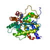
| Title | Schistosoma mansoni (Blood Fluke) Sulfotransferase/CIDD-0000773 (Compound 11g) Complex | ||||||
 Components Components | Sulfotransferase oxamniquine resistance protein | ||||||
 Keywords Keywords | TRANSFERASE / sulfotransferase / parasite / drug resistance | ||||||
| Function / homology |  Function and homology information Function and homology informationSulfotransferase, S. mansonii-type / Sulfotransferase domain / P-loop containing nucleotide triphosphate hydrolases / Rossmann fold / P-loop containing nucleoside triphosphate hydrolase / 3-Layer(aba) Sandwich / Alpha Beta Similarity search - Domain/homology | ||||||
| Biological species |   Schistosoma mansoni (invertebrata) Schistosoma mansoni (invertebrata) | ||||||
| Method |  X-RAY DIFFRACTION / MOLECULAR REPLACEMENT / Resolution: 1.444 Å X-RAY DIFFRACTION / MOLECULAR REPLACEMENT / Resolution: 1.444 Å | ||||||
 Authors Authors | Taylor, A.B. | ||||||
 Citation Citation | Journal: ACS Med Chem Lett / Year: 2018 Title: Design, Synthesis, and Characterization of Novel Small Molecules as Broad Range Antischistosomal Agents. Authors: Rugel, A. / Tarpley, R.S. / Lopez, A. / Menard, T. / Guzman, M.A. / Taylor, A.B. / Cao, X. / Kovalskyy, D. / Chevalier, F.D. / Anderson, T.J.C. / Hart, P.J. / LoVerde, P.T. / McHardy, S.F. | ||||||
| History |
|
- Structure visualization
| Structure viewer | Molecule: MolmilJmol/JSmol |
|---|
- Downloads & links
-Download
| PDBx/mmCIF format | 6mfe.cif.gz | 131.8 KB | Display | PDBx/mmCIF format |
|---|---|---|---|---|
| PDB format | pdb6mfe.ent.gz | 98.6 KB | Display | PDB format |
| PDBx/mmJSON format | 6mfe.json.gz | Tree view | PDBx/mmJSON format | |
| Others |  Other downloads Other downloads |
-Validation report
| Arichive directory | https://data.pdbj.org/pub/pdb/validation_reports/mf/6mfeftp://data.pdbj.org/pub/pdb/validation_reports/mf/6mfe | HTTPS FTP |
|---|
-Related structure data
| Related structure data |  6bdpC  6bdqC  6bdrC  6bdsC  4muaS S: Starting model for refinement C: citing same article ( |
|---|---|
| Similar structure data |




-Links
 PDBj
PDBj
- Assembly
| Deposited unit | 
| ||||||||
|---|---|---|---|---|---|---|---|---|---|
| 1 |
| ||||||||
| Unit cell |
| ||||||||
| Components on special symmetry positions |
|
-Components
| #1: Protein | Mass: 30080.547 Da / Num. of mol.: 1 Source method: isolated from a genetically manipulated source Source: (gene. exp.) Schistosoma mansoni (invertebrata) / Gene: SULT-OR, Smp_089320 / Plasmid: pAG8H / Production host:   Escherichia coli BL21 (bacteria) / Strain (production host): BL21 pLysS / References: UniProt: G4VLE5 Escherichia coli BL21 (bacteria) / Strain (production host): BL21 pLysS / References: UniProt: G4VLE5 |
|---|---|
| #2: Chemical | ChemComp-A3P / 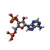 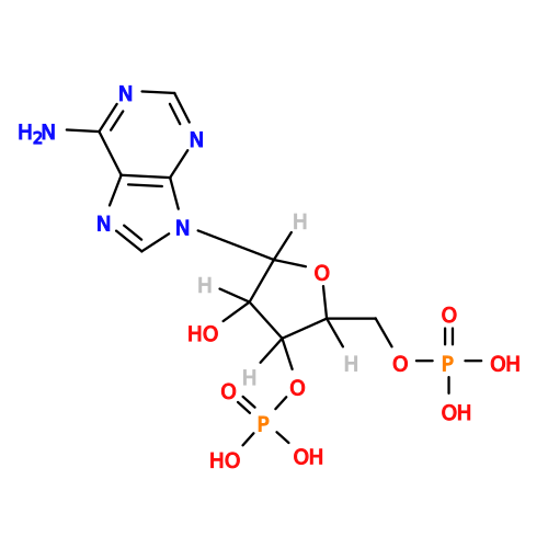 Type: RNA linking / Mass: 427.201 Da / Num. of mol.: 1 / Source method: obtained synthetically / Formula: C10H15N5O10P2 Type: RNA linking / Mass: 427.201 Da / Num. of mol.: 1 / Source method: obtained synthetically / Formula: C10H15N5O10P2 |
| #3: Chemical | ChemComp-S73 / (  Mass: 411.375 Da / Num. of mol.: 1 / Source method: obtained synthetically / Formula: C19H20F3N3O4 Mass: 411.375 Da / Num. of mol.: 1 / Source method: obtained synthetically / Formula: C19H20F3N3O4 |
| #4: Water | ChemComp-HOH /  Mass: 18.015 Da / Num. of mol.: 337 / Source method: isolated from a natural source / Formula: H2O Mass: 18.015 Da / Num. of mol.: 337 / Source method: isolated from a natural source / Formula: H2O |
| Has protein modification | Y |
-Experimental details
-Experiment
| Experiment | Method: X-RAY DIFFRACTION / Number of used crystals: 1 |
|---|
- Sample preparation
| Crystal | Density Matthews: 2.48 Å3/Da / Density % sol: 50.42 % |
|---|---|
| Crystal grow | Temperature: 295 K / Method: vapor diffusion, hanging drop / pH: 6.5 Details: 1.0 M sodium citrate, 0.1 M sodium cacodylate, pH 6.5 |
-Data collection
| Diffraction | Mean temperature: 100 K |
|---|---|
| Diffraction source | Source: ROTATING ANODE / Type: RIGAKU MICROMAX-007 HF / Wavelength: 1.54178 Å |
| Detector | Type: RIGAKU RAXIS HTC / Detector: IMAGE PLATE / Date: Jan 9, 2015 |
| Radiation | Protocol: SINGLE WAVELENGTH / Monochromatic (M) / Laue (L): M / Scattering type: x-ray |
| Radiation wavelength | Wavelength: 1.54178 Å / Relative weight: 1 |
| Reflection | Resolution: 1.44→42.66 Å / Num. obs: 52276 / % possible obs: 95.7 % / Redundancy: 6.7 % / Biso Wilson estimate: 15.1 Å2 / Rpim(I) all: 0.027 / Rsym value: 0.067 / Net I/σ(I): 17.2 |
| Reflection shell | Resolution: 1.44→1.52 Å / Redundancy: 5.9 % / Mean I/σ(I) obs: 2.1 / Num. unique obs: 6778 / Rpim(I) all: 0.351 / Rsym value: 0.818 / % possible all: 86.2 |
- Processing
| Software |
| |||||||||||||||||||||||||||||||||||||||||||||||||||||||||||||||||||||||||||||||||||||||||||||||||||||||||
|---|---|---|---|---|---|---|---|---|---|---|---|---|---|---|---|---|---|---|---|---|---|---|---|---|---|---|---|---|---|---|---|---|---|---|---|---|---|---|---|---|---|---|---|---|---|---|---|---|---|---|---|---|---|---|---|---|---|---|---|---|---|---|---|---|---|---|---|---|---|---|---|---|---|---|---|---|---|---|---|---|---|---|---|---|---|---|---|---|---|---|---|---|---|---|---|---|---|---|---|---|---|---|---|---|---|---|
| Refinement | Method to determine structure: MOLECULAR REPLACEMENT Starting model: PDB entry 4MUA Resolution: 1.444→42.656 Å / SU ML: 0.18 / Cross valid method: THROUGHOUT / σ(F): 1.35 / Phase error: 20.28
| |||||||||||||||||||||||||||||||||||||||||||||||||||||||||||||||||||||||||||||||||||||||||||||||||||||||||
| Solvent computation | Shrinkage radii: 0.9 Å / VDW probe radii: 1.11 Å | |||||||||||||||||||||||||||||||||||||||||||||||||||||||||||||||||||||||||||||||||||||||||||||||||||||||||
| Refinement step | Cycle: LAST / Resolution: 1.444→42.656 Å
| |||||||||||||||||||||||||||||||||||||||||||||||||||||||||||||||||||||||||||||||||||||||||||||||||||||||||
| Refine LS restraints |
| |||||||||||||||||||||||||||||||||||||||||||||||||||||||||||||||||||||||||||||||||||||||||||||||||||||||||
| LS refinement shell |
|
