 Movie
Movie Controller

[English] 日本語
 Yorodumi
- PDB-6uux: Schistosoma mansoni (Blood Fluke) Sulfotransferase/Hycanthone Complex -
+ Open data

- Basic information
| Entry | Database: PDB / ID: 6uux | ||||||
|---|---|---|---|---|---|---|---|
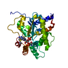
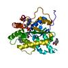
| Title | Schistosoma mansoni (Blood Fluke) Sulfotransferase/Hycanthone Complex | ||||||
 Components Components | Sulfotransferase | ||||||
 Keywords Keywords | TRANSFERASE / sulfotransferase / parasite / hycanthone / drug resistance / schistosome | ||||||
| Function / homology |  Function and homology information Function and homology information | ||||||
| Biological species |  | ||||||
| Method |  X-RAY DIFFRACTION / MOLECULAR REPLACEMENT / Resolution: 1.5 Å X-RAY DIFFRACTION / MOLECULAR REPLACEMENT / Resolution: 1.5 Å | ||||||
 Authors Authors | Taylor, A.B. | ||||||
| Funding support |  United States, 1items United States, 1items
| ||||||
 Citation Citation | Journal: Mol.Biochem.Parasitol. / Year: 2020 Title: Molecular basis for hycanthone drug action in schistosome parasites. Authors: Guzman, M. / Rugel, A. / Tarpley, R.S. / Cao, X. / McHardy, S.F. / LoVerde, P.T. / Taylor, A.B. | ||||||
| History |
|
- Structure visualization
| Structure viewer | Molecule: MolmilJmol/JSmol |
|---|
- Downloads & links
-Download
| PDBx/mmCIF format | 6uux.cif.gz | 136.7 KB | Display | PDBx/mmCIF format |
|---|---|---|---|---|
| PDB format | pdb6uux.ent.gz | 98.1 KB | Display | PDB format |
| PDBx/mmJSON format | 6uux.json.gz | Tree view | PDBx/mmJSON format | |
| Others |  Other downloads Other downloads |
-Validation report
| Arichive directory | https://data.pdbj.org/pub/pdb/validation_reports/uu/6uuxftp://data.pdbj.org/pub/pdb/validation_reports/uu/6uux | HTTPS FTP |
|---|
-Related structure data
| Related structure data |  6uuyC  5bykS S: Starting model for refinement C: citing same article ( |
|---|---|
| Similar structure data |


-Links
 PDBj
PDBj
- Assembly
| Deposited unit | 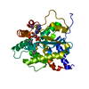
| ||||||||||||
|---|---|---|---|---|---|---|---|---|---|---|---|---|---|
| 1 |
| ||||||||||||
| Unit cell |
| ||||||||||||
| Components on special symmetry positions |
|
-Components
| #1: Protein | Mass: 30080.547 Da / Num. of mol.: 1 Source method: isolated from a genetically manipulated source Source: (gene. exp.)  References: UniProt: G4VLE5, Transferases; Transferring sulfur-containing groups; Sulfotransferases |
|---|---|
| #2: Chemical | ChemComp-A3P /   Type: RNA linking / Mass: 427.201 Da / Num. of mol.: 1 / Source method: obtained synthetically / Formula: C10H15N5O10P2 Type: RNA linking / Mass: 427.201 Da / Num. of mol.: 1 / Source method: obtained synthetically / Formula: C10H15N5O10P2 |
| #3: Chemical | ChemComp-QHM /   Mass: 356.482 Da / Num. of mol.: 1 / Source method: obtained synthetically / Formula: C20H24N2O2S / Feature type: SUBJECT OF INVESTIGATION Mass: 356.482 Da / Num. of mol.: 1 / Source method: obtained synthetically / Formula: C20H24N2O2S / Feature type: SUBJECT OF INVESTIGATION |
| #4: Water | ChemComp-HOH /  Mass: 18.015 Da / Num. of mol.: 328 / Source method: isolated from a natural source / Formula: H2O Mass: 18.015 Da / Num. of mol.: 328 / Source method: isolated from a natural source / Formula: H2O |
| Has ligand of interest | Y |
| Has protein modification | Y |
-Experimental details
-Experiment
| Experiment | Method: X-RAY DIFFRACTION / Number of used crystals: 1 |
|---|
- Sample preparation
| Crystal | Density Matthews: 2.48 Å3/Da / Density % sol: 50.34 % |
|---|---|
| Crystal grow | Temperature: 295 K / Method: vapor diffusion, sitting drop Details: 1.0 M sodium citrate, 0.1 M sodium cacodylate, pH 6.5 |
-Data collection
| Diffraction | Mean temperature: 100 K / Serial crystal experiment: N |
|---|---|
| Diffraction source | Source: ROTATING ANODE / Type: RIGAKU MICROMAX-007 HF / Wavelength: 1.54178 Å |
| Detector | Type: RIGAKU RAXIS HTC / Detector: IMAGE PLATE / Date: Aug 26, 2016 |
| Radiation | Protocol: SINGLE WAVELENGTH / Monochromatic (M) / Laue (L): M / Scattering type: x-ray |
| Radiation wavelength | Wavelength: 1.54178 Å / Relative weight: 1 |
| Reflection | Resolution: 1.5→39.3 Å / Num. obs: 48756 / % possible obs: 100 % / Redundancy: 17.7 % / Biso Wilson estimate: 18.58 Å2 / CC1/2: 0.999 / Rmerge(I) obs: 0.115 / Rpim(I) all: 0.028 / Rrim(I) all: 0.119 / Net I/σ(I): 15.1 |
| Reflection shell | Resolution: 1.5→1.58 Å / Redundancy: 10.7 % / Rmerge(I) obs: 1.429 / Mean I/σ(I) obs: 1.8 / Num. unique obs: 7017 / CC1/2: 0.382 / Rpim(I) all: 0.447 / Rrim(I) all: 1.499 / % possible all: 100 |
- Processing
| Software |
| |||||||||||||||||||||||||||||||||||||||||||||||||||||||||||||||||||||||||||||||||||||||||||||||||||||||||
|---|---|---|---|---|---|---|---|---|---|---|---|---|---|---|---|---|---|---|---|---|---|---|---|---|---|---|---|---|---|---|---|---|---|---|---|---|---|---|---|---|---|---|---|---|---|---|---|---|---|---|---|---|---|---|---|---|---|---|---|---|---|---|---|---|---|---|---|---|---|---|---|---|---|---|---|---|---|---|---|---|---|---|---|---|---|---|---|---|---|---|---|---|---|---|---|---|---|---|---|---|---|---|---|---|---|---|
| Refinement | Method to determine structure: MOLECULAR REPLACEMENT Starting model: PDB entry 5BYK Resolution: 1.5→35.29 Å / SU ML: 0.1682 / Cross valid method: THROUGHOUT / σ(F): 1.36 / Phase error: 21.2533
| |||||||||||||||||||||||||||||||||||||||||||||||||||||||||||||||||||||||||||||||||||||||||||||||||||||||||
| Solvent computation | Shrinkage radii: 0.9 Å / VDW probe radii: 1.11 Å | |||||||||||||||||||||||||||||||||||||||||||||||||||||||||||||||||||||||||||||||||||||||||||||||||||||||||
| Displacement parameters | Biso mean: 25.87 Å2 | |||||||||||||||||||||||||||||||||||||||||||||||||||||||||||||||||||||||||||||||||||||||||||||||||||||||||
| Refinement step | Cycle: LAST / Resolution: 1.5→35.29 Å
| |||||||||||||||||||||||||||||||||||||||||||||||||||||||||||||||||||||||||||||||||||||||||||||||||||||||||
| Refine LS restraints |
| |||||||||||||||||||||||||||||||||||||||||||||||||||||||||||||||||||||||||||||||||||||||||||||||||||||||||
| LS refinement shell |
|
