Movie
Movie Controller

[English] 日本語
 Yorodumi
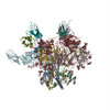
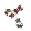
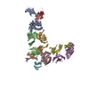
- PDB-6met: Structural basis of coreceptor recognition by HIV-1 envelope spike -
+ Open data

- Basic information
| Entry | Database: PDB / ID: 6met | |||||||||
|---|---|---|---|---|---|---|---|---|---|---|
| Title | Structural basis of coreceptor recognition by HIV-1 envelope spike | |||||||||
 Components Components |
| |||||||||
 Keywords Keywords | MEMBRANE PROTEIN / HIV coreceptor | |||||||||
| Function / homology |  Function and homology information Function and homology informationchemokine (C-C motif) ligand 5 binding / negative regulation of macrophage apoptotic process / helper T cell enhancement of adaptive immune response / interleukin-16 binding / interleukin-16 receptor activity / chemokine receptor activity / maintenance of protein location in cell / cellular response to ionomycin / response to methamphetamine hydrochloride / signaling ...chemokine (C-C motif) ligand 5 binding / negative regulation of macrophage apoptotic process / helper T cell enhancement of adaptive immune response / interleukin-16 binding / interleukin-16 receptor activity / chemokine receptor activity / maintenance of protein location in cell / cellular response to ionomycin / response to methamphetamine hydrochloride / signaling / T cell selection / phosphatidylinositol-4,5-bisphosphate phospholipase C activity / MHC class II protein binding / interleukin-15-mediated signaling pathway / cellular response to granulocyte macrophage colony-stimulating factor stimulus / positive regulation of monocyte differentiation / C-C chemokine binding / C-C chemokine receptor activity / Alpha-defensins / Nef Mediated CD4 Down-regulation / response to cholesterol / regulation of T cell activation / response to vitamin D / release of sequestered calcium ion into cytosol by sarcoplasmic reticulum / Chemokine receptors bind chemokines / Other interleukin signaling / dendritic cell chemotaxis / extracellular matrix structural constituent / T cell receptor complex / enzyme-linked receptor protein signaling pathway / Translocation of ZAP-70 to Immunological synapse / Phosphorylation of CD3 and TCR zeta chains / Interleukin-10 signaling / Generation of second messenger molecules / macrophage differentiation / immunoglobulin binding / T cell differentiation / Co-inhibition by PD-1 / positive regulation of calcium ion transport into cytosol / Binding and entry of HIV virion / positive regulation of interleukin-2 production / cellular defense response / coreceptor activity / positive regulation of T cell proliferation / positive regulation of calcium-mediated signaling / cell surface receptor protein tyrosine kinase signaling pathway / protein tyrosine kinase binding / host cell endosome membrane / cell chemotaxis / clathrin-coated endocytic vesicle membrane / calcium-mediated signaling / Vpu mediated degradation of CD4 / chemotaxis / calcium ion transport / MHC class II protein complex binding / response to estradiol / transmembrane signaling receptor activity / MAPK cascade / cell-cell signaling / Downstream TCR signaling / Cargo recognition for clathrin-mediated endocytosis / T cell receptor signaling pathway / Clathrin-mediated endocytosis / virus receptor activity / cellular response to lipopolysaccharide / positive regulation of cytosolic calcium ion concentration / signaling receptor activity / actin binding / clathrin-dependent endocytosis of virus by host cell / G alpha (i) signalling events / defense response to Gram-negative bacterium / adaptive immune response / response to ethanol / early endosome / cell surface receptor signaling pathway / cell adhesion / endosome / immune response / membrane raft / endoplasmic reticulum lumen / G protein-coupled receptor signaling pathway / inflammatory response / external side of plasma membrane / fusion of virus membrane with host endosome membrane / apoptotic process / viral envelope / symbiont entry into host cell / lipid binding / protein kinase binding / endoplasmic reticulum membrane / virion attachment to host cell / host cell plasma membrane / virion membrane / structural molecule activity / enzyme binding / cell surface / signal transduction / protein homodimerization activity / zinc ion binding / identical protein binding Similarity search - Function | |||||||||
| Biological species |   Human immunodeficiency virus 1 Human immunodeficiency virus 1 Homo sapiens (human) Homo sapiens (human) | |||||||||
| Method | ELECTRON MICROSCOPY / single particle reconstruction / cryo EM / Resolution: 4.5 Å | |||||||||
 Authors Authors | Shaik, M.M. / Chen, B. | |||||||||
| Funding support |  United States, 1items United States, 1items
| |||||||||
 Citation Citation | Journal: Nature / Year: 2019 Title: Structural basis of coreceptor recognition by HIV-1 envelope spike. Authors: Md Munan Shaik / Hanqin Peng / Jianming Lu / Sophia Rits-Volloch / Chen Xu / Maofu Liao / Bing Chen /  Abstract: HIV-1 envelope glycoprotein (Env), which consists of trimeric (gp160) cleaved to (gp120 and gp41), interacts with the primary receptor CD4 and a coreceptor (such as chemokine receptor CCR5) to fuse ...HIV-1 envelope glycoprotein (Env), which consists of trimeric (gp160) cleaved to (gp120 and gp41), interacts with the primary receptor CD4 and a coreceptor (such as chemokine receptor CCR5) to fuse viral and target-cell membranes. The gp120-coreceptor interaction has previously been proposed as the most crucial trigger for unleashing the fusogenic potential of gp41. Here we report a cryo-electron microscopy structure of a full-length gp120 in complex with soluble CD4 and unmodified human CCR5, at 3.9 Å resolution. The V3 loop of gp120 inserts into the chemokine-binding pocket formed by seven transmembrane helices of CCR5, and the N terminus of CCR5 contacts the CD4-induced bridging sheet of gp120. CCR5 induces no obvious allosteric changes in gp120 that can propagate to gp41; it does bring the Env trimer close to the target membrane. The N terminus of gp120, which is gripped by gp41 in the pre-fusion or CD4-bound Env, flips back in the CCR5-bound conformation and may irreversibly destabilize gp41 to initiate fusion. The coreceptor probably functions by stabilizing and anchoring the CD4-induced conformation of Env near the cell membrane. These results advance our understanding of HIV-1 entry into host cells and may guide the development of vaccines and therapeutic agents. | |||||||||
| History |
|
- Structure visualization
| Movie |
Movie viewer |
|---|---|
| Structure viewer | Molecule: MolmilJmol/JSmol |
- Downloads & links
-Download
| PDBx/mmCIF format | 6met.cif.gz | 240.8 KB | Display | PDBx/mmCIF format |
|---|---|---|---|---|
| PDB format | pdb6met.ent.gz | 187.8 KB | Display | PDB format |
| PDBx/mmJSON format | 6met.json.gz | Tree view | PDBx/mmJSON format | |
| Others |  Other downloads Other downloads |
-Validation report
| Arichive directory | https://data.pdbj.org/pub/pdb/validation_reports/me/6metftp://data.pdbj.org/pub/pdb/validation_reports/me/6met | HTTPS FTP |
|---|
-Related structure data
| Related structure data |  9109MC  9108C  6meoC M: map data used to model this data C: citing same article ( |
|---|---|
| Similar structure data |


-Links
 PDBj
PDBj


- Assembly
| Deposited unit | 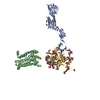
|
|---|---|
| 1 |
|
-Components
-Protein , 3 types, 3 molecules GAB
| #1: Protein | Mass: 51465.043 Da / Num. of mol.: 1 / Fragment: GP120 domain residues 29-489 Source method: isolated from a genetically manipulated source Source: (gene. exp.) Human immunodeficiency virus 1 / Gene: env / Cell line (production host): 293T / Production host: Homo sapiens (human) / References: UniProt: Q70145 |
|---|---|
| #2: Protein | Mass: 40455.477 Da / Num. of mol.: 1 Fragment: Ig-like V-type 1 and Ig-like C2-type 1,2,3 domains, residues 26-388 Source method: isolated from a genetically manipulated source Source: (gene. exp.) Homo sapiens (human) / Gene: CD4 / Cell line (production host): 293T / Production host: Homo sapiens (human) / References: UniProt: P01730 |
| #3: Protein | Mass: 36343.008 Da / Num. of mol.: 1 / Fragment: residues 1-313 Source method: isolated from a genetically manipulated source Source: (gene. exp.) Homo sapiens (human) / Gene: CCR5, CMKBR5 / Cell line (production host): 293T / Production host: Homo sapiens (human) / References: UniProt: P51681 |
-Sugars , 5 types, 17 molecules 

| #4: Polysaccharide | 2-acetamido-2-deoxy-beta-D-glucopyranose-(1-4)-2-acetamido-2-deoxy-beta-D-glucopyranose Source method: isolated from a genetically manipulated source #5: Polysaccharide | Source method: isolated from a genetically manipulated source #6: Polysaccharide | alpha-D-mannopyranose-(1-2)-alpha-D-mannopyranose-(1-3)-[alpha-D-mannopyranose-(1-6)]beta-D- ...alpha-D-mannopyranose-(1-2)-alpha-D-mannopyranose-(1-3)-[alpha-D-mannopyranose-(1-6)]beta-D-mannopyranose-(1-4)-2-acetamido-2-deoxy-beta-D-glucopyranose-(1-4)-2-acetamido-2-deoxy-beta-D-glucopyranose | Source method: isolated from a genetically manipulated source #7: Sugar | ChemComp-NAG /  Type: D-saccharide, beta linking / Mass: 221.208 Da / Num. of mol.: 7 Type: D-saccharide, beta linking / Mass: 221.208 Da / Num. of mol.: 7Source method: isolated from a genetically manipulated source Formula: C8H15NO6 #8: Sugar | ChemComp-A2G / |  Type: D-saccharide, alpha linking / Mass: 221.208 Da / Num. of mol.: 1 Type: D-saccharide, alpha linking / Mass: 221.208 Da / Num. of mol.: 1Source method: isolated from a genetically manipulated source Formula: C8H15NO6 |
|---|
-Details
| Has protein modification | Y |
|---|
-Experimental details
-Experiment
| Experiment | Method: ELECTRON MICROSCOPY |
|---|---|
| EM experiment | Aggregation state: PARTICLE / 3D reconstruction method: single particle reconstruction |
- Sample preparation
| Component |
| ||||||||||||||||||||||||||||||
|---|---|---|---|---|---|---|---|---|---|---|---|---|---|---|---|---|---|---|---|---|---|---|---|---|---|---|---|---|---|---|---|
| Molecular weight | Value: 200 MDa / Experimental value: NO | ||||||||||||||||||||||||||||||
| Source (natural) |
| ||||||||||||||||||||||||||||||
| Source (recombinant) |
| ||||||||||||||||||||||||||||||
| Buffer solution | pH: 8 Details: 100 mM Tris-HCl, pH 8.0, 150 mM NaCl, 1 mM EDTA, 0.001% LMNG (w/v), 0.025% DDM (w/v), and 0.04 % CHS (w/v). | ||||||||||||||||||||||||||||||
| Buffer component |
| ||||||||||||||||||||||||||||||
| Specimen | Conc.: 1 mg/ml / Embedding applied: NO / Shadowing applied: NO / Staining applied: NO / Vitrification applied: YES | ||||||||||||||||||||||||||||||
| Vitrification | Instrument: FEI VITROBOT MARK IV / Cryogen name: ETHANE / Humidity: 95 % / Chamber temperature: 298 K |
- Electron microscopy imaging
| Experimental equipment |  Model: Titan Krios / Image courtesy: FEI Company |
|---|---|
| Microscopy | Model: FEI TITAN KRIOS |
| Electron gun | Electron source:  FIELD EMISSION GUN / Accelerating voltage: 300 kV / Illumination mode: OTHER FIELD EMISSION GUN / Accelerating voltage: 300 kV / Illumination mode: OTHER |
| Electron lens | Mode: OTHER / Cs: 2.7 mm / Alignment procedure: BASIC |
| Specimen holder | Specimen holder model: OTHER |
| Image recording | Electron dose: 46 e/Å2 / Detector mode: SUPER-RESOLUTION / Film or detector model: GATAN K2 SUMMIT (4k x 4k) |
- Processing
| EM software |
| |||||||||||||||||||||||||
|---|---|---|---|---|---|---|---|---|---|---|---|---|---|---|---|---|---|---|---|---|---|---|---|---|---|---|
| CTF correction | Type: PHASE FLIPPING AND AMPLITUDE CORRECTION | |||||||||||||||||||||||||
| Particle selection | Num. of particles selected: 307346 | |||||||||||||||||||||||||
| Symmetry | Point symmetry: C1 (asymmetric) | |||||||||||||||||||||||||
| 3D reconstruction | Resolution: 4.5 Å / Resolution method: FSC 0.143 CUT-OFF / Num. of particles: 307346 / Num. of class averages: 1 / Symmetry type: POINT | |||||||||||||||||||||||||
| Atomic model building | B value: 200 / Protocol: RIGID BODY FIT / Space: REAL | |||||||||||||||||||||||||
| Atomic model building | 3D fitting-ID: 1 / Source name: PDB / Type: experimental model
|